
 PDF Datasheet
Human Anti-NDRG1 Antibody Product Attributes
NDRG1 Previously Observed Antibody Staining Patterns
Observed Subcellular, Organelle Specific Staining Data:
Anti-NDRG1 antibody staining is expected to be primarily localized to the cytosol and microtubules.
Observed Antibody Staining Data By Tissue Type:
Variations in NDRG1 antibody staining intensity in immunohistochemistry on tissue sections are present across different anatomical locations. An intense signal was observed in glandular cells in the adrenal gland and appendix, glial cells in the caudate nucleus and cerebral cortex, neuronal cells in the cerebral cortex, squamous epithelial cells in the cervix, uterine, glandular cells in the colon and duodenum, squamous epithelial cells in the esophagus, glandular cells in the fallopian tube and gallbladder, glial cells in the hippocampus, cells in the tubules in kidney, bile duct cells in the liver, respiratory epithelial cells in the nasopharynx, ovarian stroma cells in the ovary, islets of Langerhans in pancreas, glandular cells in the parathyroid gland, prostate, rectum and seminal vesicle, epidermal cells in the skin, glandular cells in the small intestine and stomach, cells in the seminiferous ducts in testis, squamous epithelial cells in the tonsil, urothelial cells in the urinary bladder and squamous epithelial cells in the vagina. More moderate antibody staining intensity was present in glandular cells in the adrenal gland and appendix, glial cells in the caudate nucleus and cerebral cortex, neuronal cells in the cerebral cortex, squamous epithelial cells in the cervix, uterine, glandular cells in the colon and duodenum, squamous epithelial cells in the esophagus, glandular cells in the fallopian tube and gallbladder, glial cells in the hippocampus, cells in the tubules in kidney, bile duct cells in the liver, respiratory epithelial cells in the nasopharynx, ovarian stroma cells in the ovary, islets of Langerhans in pancreas, glandular cells in the parathyroid gland, prostate, rectum and seminal vesicle, epidermal cells in the skin, glandular cells in the small intestine and stomach, cells in the seminiferous ducts in testis, squamous epithelial cells in the tonsil, urothelial cells in the urinary bladder and squamous epithelial cells in the vagina. Low, but measureable presence of NDRG1 could be seen inlymphoid tissue in appendix, glandular cells in the breast, myoepithelial cells in the breast, cells in the molecular layer in cerebellum, endothelial cells in the cerebral cortex, glandular cells in the cervix, uterine, cells in the endometrial stroma in endometrium, hepatocytes in liver, non-germinal center cells in the lymph node, exocrine glandular cells in the pancreas, smooth muscle cells in the smooth muscle, fibroblasts in mesenchymal tissue, cells in the red pulp in spleen, Leydig cells in the testis and non-germinal center cells in the tonsil. We were unable to detect NDRG1 in other tissues. Disease states, inflammation, and other physiological changes can have a substantial impact on antibody staining patterns. These measurements were all taken in tissues deemed normal or from patients without known disease.
Observed Antibody Staining Data By Tissue Disease Status:
Tissues from cancer patients, for instance, have their own distinct pattern of NDRG1 expression as measured by anti-NDRG1 antibody immunohistochemical staining. The average level of expression by tumor is summarized in the table below. The variability row represents patient to patient variability in IHC staining.
| Sample Type | breast cancer | carcinoid | cervical cancer | colorectal cancer | endometrial cancer | glioma | head and neck cancer | liver cancer | lung cancer | lymphoma | melanoma | ovarian cancer | pancreatic cancer | prostate cancer | renal cancer | skin cancer | stomach cancer | testicular cancer | thyroid cancer | urothelial cancer |
|---|---|---|---|---|---|---|---|---|---|---|---|---|---|---|---|---|---|---|---|---|
| Signal Intensity | ++ | +++ | +++ | +++ | +++ | ++ | +++ | ++ | +++ | ++ | +++ | +++ | +++ | +++ | +++ | +++ | +++ | + | + | +++ |
| NDRG1 Variability | ++ | ++ | ++ | ++ | + | ++ | ++ | +++ | ++ | ++ | ++ | ++ | ++ | + | + | ++ | ++ | ++ | ++ | + |
| NDRG1 General Information | |
|---|---|
| Alternate Names | |
| NDRG1 | |
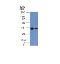
| Molecular Weight | |
| 43kDa | |
| Chromosomal Location | |
| 8q24.3 | |
| Curated Database and Bioinformatic Data | |
| Gene Symbol | NDRG1 |
| Entrez Gene ID | 10397 |
| Ensemble Gene ID | ENSG00000104419 |
| RefSeq Protein Accession(s) | NP_001128714, NP_001245361, NP_006087, XP_011515094, NP_001245362, XP_011515093 |
| RefSeq mRNA Accession(s) | NM_001258432, NM_001258433, NM_006096, XM_011516791, NM_001135242, XM_011516792 |
| RefSeq Genomic Accession(s) | NC_000008, NC_018919, NG_007943 |
| UniProt ID(s) | B3KWB2, Q8N959, Q92597 |
| UniGene ID(s) | B3KWB2, Q8N959, Q92597 |
| HGNC ID(s) | 7679 |
| Cosmic ID(s) | NDRG1 |
| KEGG Gene ID(s) | hsa:10397 |
| PharmGKB ID(s) | PA31482 |
| General Description of NDRG1. | |
| This MAb recognizes a protein of 43kDa, which is identified as N-myc downstream-regulated gene 1 protein (NDRG1). The NDRG) family is comprised of four members, NDRG1, NDRG2, NDRG3 and NDRG4, which share 57-65% homology. The NDRG1 gene is ubiquitously expressed, but it is expressed most prominently in placental membranes and prostate, kidney, small intestine and ovary tissue. NDRG1 is a direct transcriptional target gene of p53 to mediated cell death and apoptosis. NDRG1 gene expression is induced by several compounds, including nickel, and produces a protein involved in stress responses, hormone responses, cell growth and differentiation. The reduced expression of NDRG1 has been found to be associated with tumor metastasis in a variety of tumors, including cancers of the breast, colon, prostate, oral cavity and oropharynx. Reportedly, overexpression of NDRG1 in hepatocellular carcinoma is an indicator of tumor aggressiveness. | |

Reviews
There are no reviews yet.