
 PDF Datasheet
Human, Mouse, Rat, Chicken, and Pig Anti-Neurofilament Antibody Product Attributes
Neurofilament Previously Observed Antibody Staining Patterns
Observed Subcellular, Organelle Specific Staining Data:
Anti-NEFH antibody staining is expected to be primarily localized to the centrosome, nuclear bodies and nucleus.
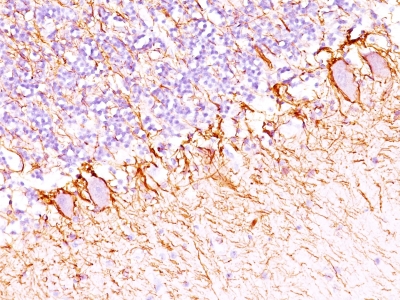
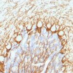
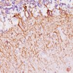
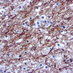
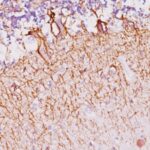
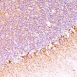
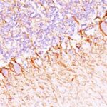
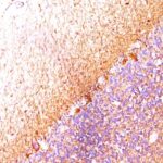
Observed Antibody Staining Data By Tissue Type:
Variations in Neurofilament antibody staining intensity in immunohistochemistry on tissue sections are present across different anatomical locations. An intense signal was observed in peripheral nerve/ganglion in colon and peripheral nerve in mesenchymal tissue. More moderate antibody staining intensity was present in peripheral nerve/ganglion in colon and peripheral nerve in mesenchymal tissue. Low, but measureable presence of Neurofilament could be seen in. We were unable to detect Neurofilament in other tissues. Disease states, inflammation, and other physiological changes can have a substantial impact on antibody staining patterns. These measurements were all taken in tissues deemed normal or from patients without known disease.
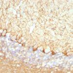
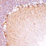
Observed Antibody Staining Data By Tissue Disease Status:
Tissues from cancer patients, for instance, have their own distinct pattern of Neurofilament expression as measured by anti-Neurofilament antibody immunohistochemical staining. The average level of expression by tumor is summarized in the table below. The variability row represents patient to patient variability in IHC staining.
| Sample Type | breast cancer | carcinoid | cervical cancer | colorectal cancer | endometrial cancer | glioma | head and neck cancer | liver cancer | lung cancer | lymphoma | melanoma | ovarian cancer | pancreatic cancer | prostate cancer | renal cancer | skin cancer | stomach cancer | testicular cancer | thyroid cancer | urothelial cancer |
|---|---|---|---|---|---|---|---|---|---|---|---|---|---|---|---|---|---|---|---|---|
| Signal Intensity | – | – | – | – | – | – | + | – | – | – | – | – | – | – | – | – | – | – | – | – |
| NEFH Variability | + | + | + | + | + | + | ++ | + | + | + | + | + | + | + | + | + | + | + | + | + |
| Neurofilament General Information | |
|---|---|
| Alternate Names | |
| NEFH, Neurofilament H, NF2, anti-NEFH antibody, anti-Neurofilament H antibody, anti-NF2 antibody | |
| Molecular Weight | |
| 200kDa | |
| Chromosomal Location | |
| 22q12.2 | |
| Curated Database and Bioinformatic Data | |
| Gene Symbol | NEFH |
| Entrez Gene ID | 4744 |
| Ensemble Gene ID | ENSG00000100285 |
| RefSeq Protein Accession(s) | XP_011528502, NP_066554 |
| RefSeq mRNA Accession(s) | NM_021076, XM_011530200 |
| RefSeq Genomic Accession(s) | NG_008404, NC_018933, NC_000022 |
| UniProt ID(s) | P12036 |
| UniGene ID(s) | P12036 |
| HGNC ID(s) | 7737 |
| Cosmic ID(s) | NEFH |
| KEGG Gene ID(s) | hsa:4744 |
| PharmGKB ID(s) | PA31540 |
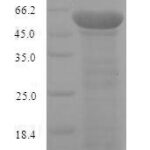
| General Description of Neurofilament. | |
| This MAb reacts with a 200kDa protein, identified as heavy sub-unit of neurofilaments (NF-H). Neurofilaments make up the main structural elements of axons, dendrites, are found in neurons, peripheral nerves,, sympathetic ganglion cells. Neurofilaments consist of three major subunits with molecular weights of 68kDa (NF-L), 160kDa (NF-M), 200kDa (NF-H). Anti-neurofilament stains a number of neural, neuroendocrine,, endocrine tumors. Neuromas, ganglioneuromas, gangliogliomas, ganglioneuroblastomas,, neuroblastomas stain positively for anti-neurofilament. Neurofilaments are also present in paragangliomas as well as adrenal, extra-adrenal pheochromocytomas. Carcinoids, neuroendocrine carcinomas of the skin,, oat cell carcinomas of the lung also express neurofilament. | |

Reviews
There are no reviews yet.