

 PDF Datasheet
Human Anti-Nuclear Mitotic Apparatus Protein Antibody Product Attributes
Nuclear Mitotic Apparatus Protein Previously Observed Antibody Staining Patterns
Observed Subcellular, Organelle Specific Staining Data:
Anti-NUMA1 antibody staining is expected to be primarily localized to the nucleoplasm.
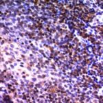
Observed Antibody Staining Data By Tissue Type:
Variations in Nuclear Mitotic Apparatus Protein antibody staining intensity in immunohistochemistry on tissue sections are present across different anatomical locations. An intense signal was observed in glandular cells in the adrenal gland and appendix, hematopoietic cells in the bone marrow, glandular cells in the breast, myoepithelial cells in the breast, respiratory epithelial cells in the bronchus, glial cells in the caudate nucleus, neuronal cells in the caudate nucleus, cells in the granular layer in cerebellum, cells in the molecular layer in cerebellum, Purkinje cells in the cerebellum, glial cells in the cerebral cortex, neuronal cells in the cerebral cortex, glandular cells in the cervix, uterine, squamous epithelial cells in the cervix, uterine, endothelial cells in the colon, glandular cells in the colon, peripheral nerve/ganglion in colon, glandular cells in the duodenum, cells in the endometrial stroma in endometrium, glandular cells in the endometrium and epididymis, squamous epithelial cells in the esophagus, glandular cells in the fallopian tube and gallbladder, myocytes in heart muscle, glial cells in the hippocampus, neuronal cells in the hippocampus, cells in the glomeruli in kidney, cells in the tubules in kidney, macrophages in lung, pneumocytes in lung, non-germinal center cells in the lymph node, respiratory epithelial cells in the nasopharynx, squamous epithelial cells in the oral mucosa, ovarian stroma cells in the ovary, exocrine glandular cells in the pancreas, islets of Langerhans in pancreas, glandular cells in the parathyroid gland, decidual cells in the placenta, trophoblastic cells in the placenta, glandular cells in the prostate, rectum, salivary gland and seminal vesicle, myocytes in skeletal muscle, fibroblasts in skin, keratinocytes in skin, Langerhans in skin, melanocytes in skin, epidermal cells in the skin, glandular cells in the small intestine, smooth muscle cells in the smooth muscle, adipocytes in mesenchymal tissue, chondrocytes in mesenchymal tissue, fibroblasts in mesenchymal tissue, peripheral nerve in mesenchymal tissue, cells in the red pulp in spleen, cells in the white pulp in spleen, glandular cells in the stomach, cells in the seminiferous ducts in testis, Leydig cells in the testis, glandular cells in the thyroid gland, non-germinal center cells in the tonsil, squamous epithelial cells in the tonsil, urothelial cells in the urinary bladder and squamous epithelial cells in the vagina. More moderate antibody staining intensity was present in glandular cells in the adrenal gland and appendix, hematopoietic cells in the bone marrow, glandular cells in the breast, myoepithelial cells in the breast, respiratory epithelial cells in the bronchus, glial cells in the caudate nucleus, neuronal cells in the caudate nucleus, cells in the granular layer in cerebellum, cells in the molecular layer in cerebellum, Purkinje cells in the cerebellum, glial cells in the cerebral cortex, neuronal cells in the cerebral cortex, glandular cells in the cervix, uterine, squamous epithelial cells in the cervix, uterine, endothelial cells in the colon, glandular cells in the colon, peripheral nerve/ganglion in colon, glandular cells in the duodenum, cells in the endometrial stroma in endometrium, glandular cells in the endometrium and epididymis, squamous epithelial cells in the esophagus, glandular cells in the fallopian tube and gallbladder, myocytes in heart muscle, glial cells in the hippocampus, neuronal cells in the hippocampus, cells in the glomeruli in kidney, cells in the tubules in kidney, macrophages in lung, pneumocytes in lung, non-germinal center cells in the lymph node, respiratory epithelial cells in the nasopharynx, squamous epithelial cells in the oral mucosa, ovarian stroma cells in the ovary, exocrine glandular cells in the pancreas, islets of Langerhans in pancreas, glandular cells in the parathyroid gland, decidual cells in the placenta, trophoblastic cells in the placenta, glandular cells in the prostate, rectum, salivary gland and seminal vesicle, myocytes in skeletal muscle, fibroblasts in skin, keratinocytes in skin, Langerhans in skin, melanocytes in skin, epidermal cells in the skin, glandular cells in the small intestine, smooth muscle cells in the smooth muscle, adipocytes in mesenchymal tissue, chondrocytes in mesenchymal tissue, fibroblasts in mesenchymal tissue, peripheral nerve in mesenchymal tissue, cells in the red pulp in spleen, cells in the white pulp in spleen, glandular cells in the stomach, cells in the seminiferous ducts in testis, Leydig cells in the testis, glandular cells in the thyroid gland, non-germinal center cells in the tonsil, squamous epithelial cells in the tonsil, urothelial cells in the urinary bladder and squamous epithelial cells in the vagina. Low, but measureable presence of Nuclear Mitotic Apparatus Protein could be seen inhepatocytes in liver. We were unable to detect Nuclear Mitotic Apparatus Protein in other tissues. Disease states, inflammation, and other physiological changes can have a substantial impact on antibody staining patterns. These measurements were all taken in tissues deemed normal or from patients without known disease.
Observed Antibody Staining Data By Tissue Disease Status:
Tissues from cancer patients, for instance, have their own distinct pattern of Nuclear Mitotic Apparatus Protein expression as measured by anti-Nuclear Mitotic Apparatus Protein antibody immunohistochemical staining. The average level of expression by tumor is summarized in the table below. The variability row represents patient to patient variability in IHC staining.
| Sample Type | breast cancer | carcinoid | cervical cancer | colorectal cancer | endometrial cancer | glioma | head and neck cancer | liver cancer | lung cancer | lymphoma | melanoma | ovarian cancer | pancreatic cancer | prostate cancer | renal cancer | skin cancer | stomach cancer | testicular cancer | thyroid cancer | urothelial cancer |
|---|---|---|---|---|---|---|---|---|---|---|---|---|---|---|---|---|---|---|---|---|
| Signal Intensity | +++ | +++ | ++ | ++ | +++ | +++ | +++ | ++ | +++ | ++ | +++ | +++ | +++ | +++ | ++ | +++ | ++ | ++ | +++ | +++ |
| NUMA1 Variability | + | ++ | ++ | ++ | ++ | + | ++ | ++ | ++ | ++ | ++ | + | ++ | + | ++ | ++ | ++ | ++ | + | ++ |
| Nuclear Mitotic Apparatus Protein General Information | |
|---|---|
| Alternate Names | |
| Nuclear mitotic apparatus protein 1, NUMA1 | |
| Molecular Weight | |
| 228kDa | |
| Chromosomal Location | |
| 11q13 | |
| Curated Database and Bioinformatic Data | |
| Gene Symbol | NUMA1 |
| Entrez Gene ID | 4926 |
| Ensemble Gene ID | ENSG00000137497 |
| RefSeq Protein Accession(s) | XP_006718627, XP_011543356, XP_011543366, XP_016873319, XP_011543362, XP_011543363, XP_011543365, XP_011543361, XP_011543368, XP_011543359, XP_011543358, XP_011543364, NP_001273490, XP_011543357, XP_011543360, XP_011543367, XP_016873320, NP_006176 |
| RefSeq mRNA Accession(s) | XM_011545059, XM_011545066, XM_017017831, NM_001286561, XM_011545054, XM_011545060, XM_011545064, XM_011545062, XM_011545065, XM_017017830, NR_104476, XM_011545063, XM_011545055, XM_011545061, NM_006185, XM_011545057, XM_011545058, XM_006718564, XM_011545056 |
| RefSeq Genomic Accession(s) | NC_018922, NC_000011 |
| UniProt ID(s) | A0A024R5M9, Q4LE64, Q14980, Q3SYK8 |
| UniGene ID(s) | A0A024R5M9, Q4LE64, Q14980, Q3SYK8 |
| HGNC ID(s) | 8059 |
| Cosmic ID(s) | NUMA1 |
| KEGG Gene ID(s) | hsa:4926 |
| PharmGKB ID(s) | PA31844 |
| General Description of Nuclear Mitotic Apparatus Protein. | |
| Recognizes a phosphorylated protein of 228kDa, identified as nuclear mitotic apparatus protein (NuMA). Its epitope is resistant to phosphatases. NuMA is intra-nuclear protein, present in nucleus during interphase. At the onset of mitosis, it redistributes from the nucleus to two centrosomal structures that later will become part of the mitotic spindle pole. After anaphase, the protein redistributes from the spindle polar region into reforming nucleus. NuMA is an essential protein during mitosis for the terminal phases of chromosome separation,/or nuclear reassembly. Recently a study shows that NuMA is cleaved to a 180 to 200kDa during apoptosis. Chromosomal translocation of this gene with the RARA (retinoic acid receptor, alpha) gene on chromosome 17 has been detected in patients with acute promyelocytic leukemia. | |
There are no reviews yet.