
 PDF Datasheet
Human, Monkey, Mouse, and Rat Anti-p27Kip1 Antibody Product Attributes
p27Kip1 Previously Observed Antibody Staining Patterns
Observed Subcellular, Organelle Specific Staining Data:
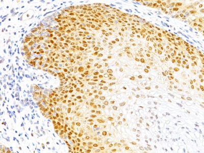
Anti-CDKN1B antibody staining is expected to be primarily localized to the nucleus and vesicles. There is variability in either the signal strength or the localization of signal in nucleus from cell to cell.
Observed Antibody Staining Data By Tissue Type:
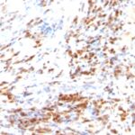
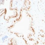
Variations in p27Kip1 antibody staining intensity in immunohistochemistry on tissue sections are present across different anatomical locations. An intense signal was observed in cells in the glomeruli in kidney, cells in the granular layer in cerebellum, follicle cells in the ovary, glandular cells in the cervix, uterine and fallopian tube, hematopoietic cells in the bone marrow, neuronal cells in the caudate nucleus and hippocampus, non-germinal center cells in the tonsil, ovarian stroma cells in the ovary, pneumocytes in lung and Purkinje cells in the cerebellum. More moderate antibody staining intensity was present in cells in the glomeruli in kidney, cells in the granular layer in cerebellum, follicle cells in the ovary, glandular cells in the cervix, uterine and fallopian tube, hematopoietic cells in the bone marrow, neuronal cells in the caudate nucleus and hippocampus, non-germinal center cells in the tonsil, ovarian stroma cells in the ovary, pneumocytes in lung and Purkinje cells in the cerebellum. Low, but measureable presence of p27Kip1 could be seen inadipocytes in breast and mesenchymal tissue, cells in the red pulp in spleen, cells in the seminiferous ducts in testis, cells in the tubules in kidney, cells in the white pulp in spleen, decidual cells in the placenta, endothelial cells in the cerebral cortex and colon, exocrine glandular cells in the pancreas, fibroblasts in mesenchymal tissue, germinal center cells in the lymph node and tonsil, glandular cells in the appendix, breast, colon, duodenum, epididymis, parathyroid gland, rectum, seminal vesicle, small intestine, stomach and thyroid gland, hepatocytes in liver, Langerhans in skin, Leydig cells in the testis, macrophages in lung, myocytes in heart muscle, myoepithelial cells in the breast, neuropil in cerebral cortex, respiratory epithelial cells in the bronchus, smooth muscle cells in the smooth muscle and squamous epithelial cells in the esophagus. We were unable to detect p27Kip1 in other tissues. Disease states, inflammation, and other physiological changes can have a substantial impact on antibody staining patterns. These measurements were all taken in tissues deemed normal or from patients without known disease.
Observed Antibody Staining Data By Tissue Disease Status:
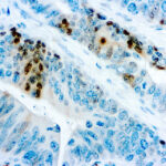
Tissues from cancer patients, for instance, have their own distinct pattern of p27Kip1 expression as measured by anti-p27Kip1 antibody immunohistochemical staining. The average level of expression by tumor is summarized in the table below. The variability row represents patient to patient variability in IHC staining.
| Sample Type | breast cancer | carcinoid | cervical cancer | colorectal cancer | endometrial cancer | glioma | head and neck cancer | liver cancer | lung cancer | lymphoma | melanoma | ovarian cancer | pancreatic cancer | prostate cancer | renal cancer | skin cancer | stomach cancer | testicular cancer | thyroid cancer | urothelial cancer |
|---|---|---|---|---|---|---|---|---|---|---|---|---|---|---|---|---|---|---|---|---|
| Signal Intensity | + | + | – | + | – | ++ | ++ | – | + | – | + | – | – | + | – | – | + | – | ++ | – |
| CDKN1B Variability | ++ | +++ | ++ | ++ | ++ | ++ | ++ | ++ | ++ | + | ++ | ++ | ++ | ++ | + | + | ++ | + | ++ | + |
| p27Kip1 General Information | |
|---|---|
| Alternate Names | |
| Cyclin-dependent kinase inhibitor 1B, p27Kip1, CDKN1B, p27Kip1, p27 Kip1 | |
| Molecular Weight | |
| 25-26kDa | |
| Chromosomal Location | |
| 12p13.1 | |
| Curated Database and Bioinformatic Data | |
| Gene Symbol | CDKN1B |
| Entrez Gene ID | 1027 |
| Ensemble Gene ID | ENSG00000111276 |
| RefSeq Protein Accession(s) | NP_004055 |
| RefSeq mRNA Accession(s) | NM_004064 |
| RefSeq Genomic Accession(s) | NG_016341, NC_018923, NC_000012 |
| UniProt ID(s) | P46527, Q6I9V6 |
| UniGene ID(s) | P46527, Q6I9V6 |
| HGNC ID(s) | 1785 |
| Cosmic ID(s) | CDKN1B |
| KEGG Gene ID(s) | hsa:1027 |
| PharmGKB ID(s) | PA105 |
| General Description of p27Kip1. | |
| Recognizes a 27kDa protein, identified as the p27Kip1, a cell cycle regulatory mitotic inhibitor. Its epitope spans between aa 83-204 of p27. It is highly specific, shows no cross-reaction with other related mitotic inhibitors. p27Kip1 functions as a negative regulator of G1 progression, has been proposed to function as a possible mediator of TGF- induced G1 arrest. p27Kip1 is a candidate tumor suppressor gene. This MAb co-precipitates cdk4 in complex p27Kip1, is excellent for staining of formalin-fixed tissues. | |
Reviews
There are no reviews yet.