

 PDF Datasheet
Human Anti-PAX6 Antibody Product Attributes
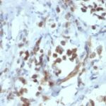
PAX6 Previously Observed Antibody Staining Patterns
Observed Subcellular, Organelle Specific Staining Data:
Anti-PAX6 antibody staining is expected to be primarily localized to the nucleoplasm.
Observed Antibody Staining Data By Tissue Type:
Variations in PAX6 antibody staining intensity in immunohistochemistry on tissue sections are present across different anatomical locations. An intense signal was observed in cells in the granular layer in cerebellum and islets of Langerhans in pancreas. More moderate antibody staining intensity was present in cells in the granular layer in cerebellum and islets of Langerhans in pancreas. Low, but measureable presence of PAX6 could be seen inglandular cells in the appendix, colon, small intestine, stomach. We were unable to detect PAX6 in other tissues. Disease states, inflammation, and other physiological changes can have a substantial impact on antibody staining patterns. These measurements were all taken in tissues deemed normal or from patients without known disease.
Observed Antibody Staining Data By Tissue Disease Status:
Tissues from cancer patients, for instance, have their own distinct pattern of PAX6 expression as measured by anti-PAX6 antibody immunohistochemical staining. The average level of expression by tumor is summarized in the table below. The variability row represents patient to patient variability in IHC staining.
| Sample Type | breast cancer | carcinoid | cervical cancer | colorectal cancer | endometrial cancer | glioma | head and neck cancer | liver cancer | lung cancer | lymphoma | melanoma | ovarian cancer | pancreatic cancer | prostate cancer | renal cancer | skin cancer | stomach cancer | testicular cancer | thyroid cancer | urothelial cancer |
|---|---|---|---|---|---|---|---|---|---|---|---|---|---|---|---|---|---|---|---|---|
| Signal Intensity | – | – | – | – | – | – | – | – | – | – | – | – | – | – | – | – | – | – | – | – |
| PAX6 Variability | + | ++ | ++ | + | + | ++ | + | + | + | + | + | + | + | + | + | ++ | + | + | + | + |
| PAX6 General Information | |
|---|---|
| Alternate Names | |
| Paired box protein Pax-6 aniridia type II protein, PAX6 | |
| Molecular Weight | |
| 47kDa | |
| Chromosomal Location | |
| 11p13 | |
| Curated Database and Bioinformatic Data | |
| Gene Symbol | PAX6 |
| Entrez Gene ID | 5080 |
| Ensemble Gene ID | ENSG00000007372 |
| RefSeq Protein Accession(s) | NP_001121084, NP_001595, NP_001245394, NP_001297087, NP_001297088, NP_001245391, NP_001245392, NP_001297089, NP_001297090, NP_001245393, NP_000271 |
| RefSeq mRNA Accession(s) | NM_001127612, NM_001258462, NM_001258463, NM_001310160, NM_001310161, NM_001258465, NM_000280, NM_001310158, NM_001604 NM_001310159, NM_001258464 |
| RefSeq Genomic Accession(s) | NG_008679, NC_000011, NC_018922 |
| UniProt ID(s) | A0A1W2PRA8, P26367, F1T0F8, Q66SS1, D1KF47 |
| UniGene ID(s) | A0A1W2PRA8, P26367, F1T0F8, Q66SS1, D1KF47 |
| HGNC ID(s) | 8620 |
| Cosmic ID(s) | PAX6 |
| KEGG Gene ID(s) | hsa:5080 |
| PharmGKB ID(s) | PA32960 |
| General Description of PAX6. | |
| Pax genes contain paired domains with strong homology to genes in Drosophila, which are involved in programming early development. Lesions in the Pax-6 gene account for most cases of aniridia, a congenital malformation of the eye, chiefly characterized by iris hypoplasia, which can cause blindness. Pax-6 is involved in other anterior segment malformations besides aniridia, such as Peters anomaly, a major error in the embryonic development of the eye with corneal clouding with variable iridolenticulocorneal adhesions. The Pax-6 gene encodes a transcriptional regulator that recognizes target genes through its paired-type DNA-binding domain. The paired domain is composed of two distinct DNA-binding subdomains, the amino-terminal subdomain, the carboxy-terminal subdomain, which bind respective consensus DNA sequences. The human Pax-6 gene produces two alternatively spliced isoforms that have the distinct structure of the paired domain. | |
There are no reviews yet.