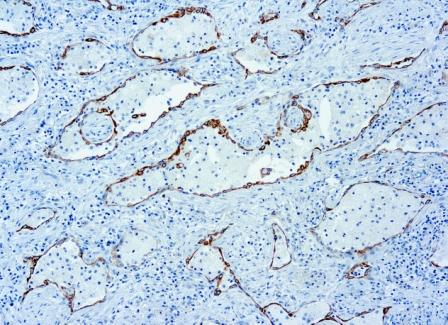
Antibody (Suitable for clinical applications)
| Specification | Recommendation |
|---|---|
| Recommended Dilution (Conc) | 1:50-1:100 |
| Pretreatment | EDTA Buffer pH 8.0 |
| Incubation Parameters | 30 min at Room Temperature |
Prior to use, inspect vial for the presence of any precipitate or other unusual physical properties. These can indicate that the antibody has degraded and is no longer suitable for patient samples. Please run positive and negative controls simultaneously with all patient samples to account and control for errors in laboratory procedure. Use of methods or materials not recommended by enQuire Bio including change to dilution range and detection system should be routinely validated by the user.
PD1 Information for Pathologists
Summary:
Also known as CD279. Coinhibitory receptor of lymphocytes / other immune system cells. Controls lymphocyte activation by providing negative signals in conjunction with signals from lymphocyte antigen receptors (Future Oncol 2011;7:929). Expressed by germinal center associated helper T cells; inhibits T cell activity (Hum Pathol 2009;40:1715). Expressed by CD8+ T cells, associated with CD8 activation (AIDS Res Hum Retroviruses 2012;28:465).
Common Uses By Pathologists:
Differentiate primary cutaneous CD4 small / medium sized pleomorphic T cell lymphoma and cutaneous pseudo-T cell lymphomas (PD-1 positive) from other cutaneous T cell lymphomas (usually PD-1 negative, Am J Surg Pathol 2012;36:109). PD-1+ rosettes of T cells around neoplastic cells is relatively specific for nodular lymphocyte predominant Hodgkin lymphoma (Hum Pathol 2009;40:1715). Microscopic (histologic) images Images hosted on PathOut server:. Contributed by GenomeMe:.
| PD1 General Information | |
|---|---|
| Alternate Names | |
| Molecular Weight | |
| 31.6 kDa | |
| Chromosomal Location | |
| q37.3 [chr: 2] [chr_start: 241849884] [chr_end: 241858894] [strand: -1]; [chr: CHR_HSCHR2_3_CTG15] [chr_start: 241849881] [chr_end: 241858908] [strand: -1] | |
| Curated Database and Bioinformatic Data | |
| Gene Symbol | PDCD1 |
| Entrez Gene ID | 5133 |
| RefSeq Protein Accession(s) | NP_005009 |
| RefSeq mRNA Accession(s) | ; NM_005018; XM_006712573; XM_017004293 |
| RefSeq Genomic Accession(s) | NT_187527; NC_000002; NG_012110 |
| UniProt ID(s) | Q15116 |
| PharmGKB ID(s) | PA33110 |
| KEGG Gene ID(s) | hsa:5133 |
| Associated Diseases (KEGG IDs) | Systemic lupus erythematosus 2 (SLEB2) [MIM:605218]: A chronic, relapsing, inflammatory, and often febrile multisystemic disorder of connective tissue, characterized principally by involvement of the skin, joints, kidneys and serosal membranes. It is of unknown etiology, but is thought to represent a failure of the regulatory mechanisms of the autoimmune system. The disease is marked by a wide range of system dysfunctions, an elevated erythrocyte sedimentation rate, and the formation of LE cells in the blood or bone marrow. {ECO:0000269|PubMed:12402038}. Disease susceptibility is associated with variations affecting the gene represented in this entry. |
| General Description of PD1 . | |
| PD1, or CD279/PDCD1 (programmed cell death-1 protein), is a type I transmembrane receptor expressed on activated T-cells, B-cells, and myeloid cells. Engagement of PD1 by its ligands PD-L1 or PD-L2 transduces a signal that inhibits T-cell proliferation, cytokine production, and cytolytic function. Anti-PDCD1 is a marker of angioimmunoblastic lymphoma.also pro¬duce responses in nonimmunogenic cancers such as non–small-cell lung and colon cancers, broadening their scope beyond classic immunogenic tumors like melanoma and renal cell cancer. | |


There are no reviews yet.