Antibody (Suitable for clinical applications)
| Specification | Recommendation |
|---|---|
| Recommended Dilution (Conc) | 1:50-1:100 |
| Pretreatment | EDTA Buffer pH 8.0 |
| Incubation Parameters | 30 min at Room Temperature |
Prior to use, inspect vial for the presence of any precipitate or other unusual physical properties. These can indicate that the antibody has degraded and is no longer suitable for patient samples. Please run positive and negative controls simultaneously with all patient samples to account and control for errors in laboratory procedure. Use of methods or materials not recommended by enQuire Bio including change to dilution range and detection system should be routinely validated by the user.
Phosphohistone H3 Information for Pathologists
Summary:
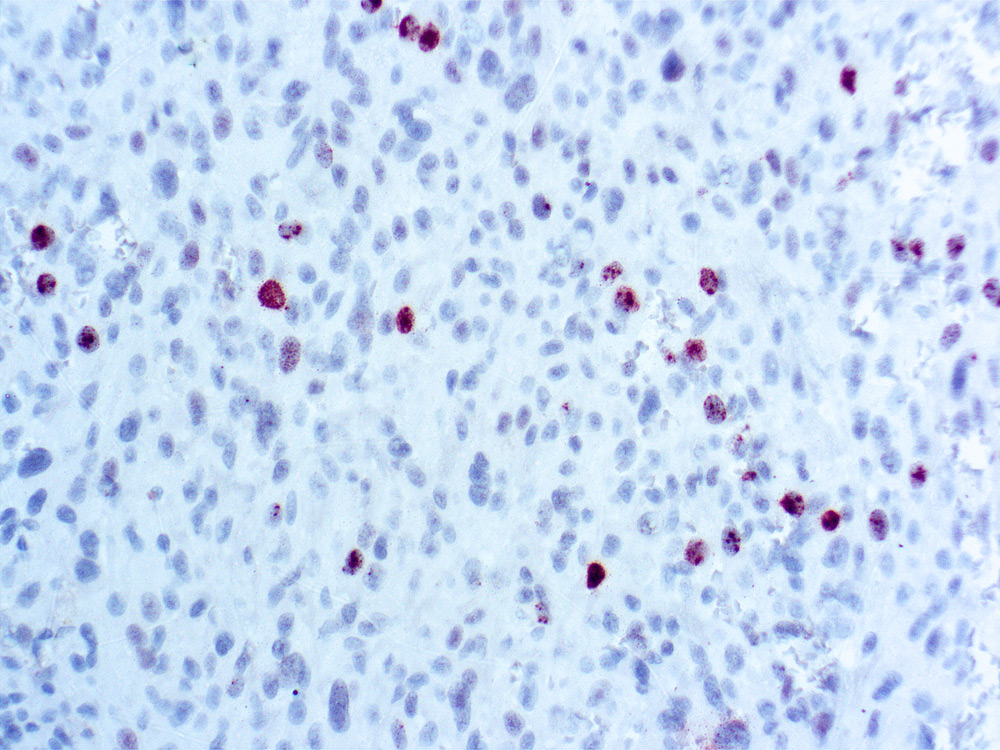
Core histone protein that is major constituent of chromatin; marker of cells in late G2 and M phase. Interpretation Nuclear stain. Count phosphohistone H3+ objects (nuclei and mitoses) in 10 adjacent fields with 40x objective, in invasive epithelial areas with highest phosphohistone H3 staining. Ignore nuclei with fine granular staining.
Common Uses By Pathologists:
Identify mitotic activity in tumor cells; use for counting mitotic figures. Strong prognostic value in lymph node negative breast cancer patients Mod Pathol 2007;20:1307, Mod Pathol 2011;24:502). Strong prognostic value (Cell Oncol 2009;31:261). Prognostic value in meningiomas (Am J Clin Pathol 2007;128:118, Am J Surg Pathol 2004;28:1532). Microscopic (histologic) images
| Phosphohistone H3 General Information | |
|---|---|
| Alternate Names | |
| Molecular Weight | |
| 15.4 kDa | |
| Curated Database and Bioinformatic Data | |
| Gene Symbol | HHT2; HHT1 |
| UniProt ID(s) | P69150 |
| General Description of Phosphohistone H3 . | |
| Phosphohistone H3 (PHH3) is a core histone protein, which together with other histones, forms the major protein constituents of the chromatin in eukaryotic cells. In mammalian cells, phosphohistone H3 is negligible during interphase but reaches a maximum for chromatin condensation during mitosis.Immunohistochemical studies showed anti-PHH3 specifically detected the core protein histone H3 only when phosphorylated at serine 10 or serine 28. Studies have also revealed no phosphorylation on the histone H3 during apoptosis.PHH3 can serve as a mitotic marker to separate mitotic figures from apoptotic bodies and karyorrhectic debris, which may be a very useful tool in diagnosis of tumor grades, especially in CNS, skin, gyn., soft tissue, and GIST. | |


Reviews
There are no reviews yet.