
 PDF Datasheet
Human and Cynomolgus Monkey Anti-pS2 / pNR-2 / Trefoil Factor 1 Antibody Product Attributes
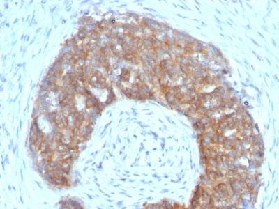
pS2 / pNR-2 / Trefoil Factor 1 Previously Observed Antibody Staining Patterns
Observed Antibody Staining Data By Tissue Type:
Variations in pS2 / pNR-2 / Trefoil Factor 1 antibody staining intensity in immunohistochemistry on tissue sections are present across different anatomical locations. An intense signal was observed in glandular cells in the stomach. More moderate antibody staining intensity was present in glandular cells in the stomach. Low, but measureable presence of pS2 / pNR-2 / Trefoil Factor 1 could be seen inglandular cells in the appendix, colon, duodenum, gallbladder, rectum, small intestine. We were unable to detect pS2 / pNR-2 / Trefoil Factor 1 in other tissues. Disease states, inflammation, and other physiological changes can have a substantial impact on antibody staining patterns. These measurements were all taken in tissues deemed normal or from patients without known disease.
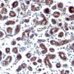
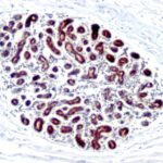
Observed Antibody Staining Data By Tissue Disease Status:
Tissues from cancer patients, for instance, have their own distinct pattern of pS2 / pNR-2 / Trefoil Factor 1 expression as measured by anti-pS2 / pNR-2 / Trefoil Factor 1 antibody immunohistochemical staining. The average level of expression by tumor is summarized in the table below. The variability row represents patient to patient variability in IHC staining.
| Sample Type | breast cancer | carcinoid | cervical cancer | colorectal cancer | endometrial cancer | glioma | head and neck cancer | liver cancer | lung cancer | lymphoma | melanoma | ovarian cancer | pancreatic cancer | prostate cancer | renal cancer | skin cancer | stomach cancer | testicular cancer | thyroid cancer | urothelial cancer |
|---|---|---|---|---|---|---|---|---|---|---|---|---|---|---|---|---|---|---|---|---|
| Signal Intensity | – | – | – | – | – | – | – | – | – | – | – | – | – | – | – | – | – | – | – | – |
| TFF1 Variability | ++ | ++ | + | + | + | + | + | + | + | + | + | ++ | ++ | + | + | + | + | + | + | + |
| pS2 / pNR-2 / Trefoil Factor 1 General Information | |
|---|---|
| Alternate Names | |
| Trefoil factor 1, TFF1 | |
| Molecular Weight | |
| 6.5kDa | |
| Chromosomal Location | |
| 16p11.2 | |
| Curated Database and Bioinformatic Data | |
| Gene Symbol | TFF1 |
| Entrez Gene ID | 7031 |
| Ensemble Gene ID | ENSG00000160182 |
| RefSeq Protein Accession(s) | NP_003216 |
| RefSeq mRNA Accession(s) | NM_003225 |
| RefSeq Genomic Accession(s) | NC_000021, NC_018932 |
| UniProt ID(s) | P04155 |
| UniGene ID(s) | P04155 |
| HGNC ID(s) | 11755 |
| Cosmic ID(s) | TFF1 |
| KEGG Gene ID(s) | hsa:7031 |
| PharmGKB ID(s) | PA36470 |
| General Description of pS2 / pNR-2 / Trefoil Factor 1. | |
| It recognizes a polypeptide of 6.5kDa, identified as pS2 estrogen-regulated protein. Its epitope is localized between aa57-84 of human pS2 protein. pS2 is a trefoil peptide. Trefoil peptides are protease resistant molecules secreted throughout the gut that play a role in mucosal healing. These peptides contain three intra-chain disulfide bonds, forming the trefoil motif, or P-domain. pS2 is known to form dimers, this dimerization is thought to play a role in its protective, healing properties. About 60% of breast carcinomas are positive for pS2. Staining is cytoplasmic, often with localization to the Golgi apparatus. pS2 is shown to be localized in normal stomach mucosa, gastric fluid, goblet cells in the colon, small intestine,, in ulcerations of the gastrointestinal tract. Several studies have shown that pS2 is primarily expressed in estrogen receptor-positive breast tumors, it may define a subset of estrogen-dependent tumors that displays an increased likelihood of response to endocrine therapy. | |

Reviews
There are no reviews yet.