
 PDF Datasheet
Human and Horse Anti-Renal Cell Carcinoma / gp200 Antibody Product Attributes
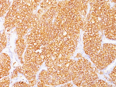
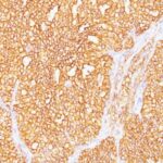
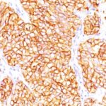
Renal Cell Carcinoma / gp200 Previously Observed Antibody Staining Patterns
Observed Subcellular, Organelle Specific Staining Data:
Anti-CA9 antibody staining is expected to be primarily localized to the plasma membrane and cytosol.
Observed Antibody Staining Data By Tissue Type:
Variations in Renal Cell Carcinoma / gp200 antibody staining intensity in immunohistochemistry on tissue sections are present across different anatomical locations. An intense signal was observed in glandular cells in the gallbladder, stomach. More moderate antibody staining intensity was present in glandular cells in the gallbladder, stomach. Low, but measureable presence of Renal Cell Carcinoma / gp200 could be seen in. We were unable to detect Renal Cell Carcinoma / gp200 in other tissues. Disease states, inflammation, and other physiological changes can have a substantial impact on antibody staining patterns. These measurements were all taken in tissues deemed normal or from patients without known disease.
Observed Antibody Staining Data By Tissue Disease Status:
Tissues from cancer patients, for instance, have their own distinct pattern of Renal Cell Carcinoma / gp200 expression as measured by anti-Renal Cell Carcinoma / gp200 antibody immunohistochemical staining. The average level of expression by tumor is summarized in the table below. The variability row represents patient to patient variability in IHC staining.
| Sample Type | breast cancer | carcinoid | cervical cancer | colorectal cancer | endometrial cancer | glioma | head and neck cancer | liver cancer | lung cancer | lymphoma | melanoma | ovarian cancer | pancreatic cancer | prostate cancer | renal cancer | skin cancer | stomach cancer | testicular cancer | thyroid cancer | urothelial cancer |
|---|---|---|---|---|---|---|---|---|---|---|---|---|---|---|---|---|---|---|---|---|
| Signal Intensity | – | – | – | – | – | – | – | – | – | – | – | – | – | – | – | – | – | – | – | – |
| CA9 Variability | + | + | + | + | + | + | + | + | + | + | + | + | + | + | ++ | + | ++ | + | + | ++ |
| Renal Cell Carcinoma / gp200 General Information | |
|---|---|
| Alternate Names | |
| Carbonic anhydrase 9, CA9, CAIX | |
| Molecular Weight | |
| 200kDa | |
| Chromosomal Location | |
| 9p13.3 | |
| Curated Database and Bioinformatic Data | |
| Gene Symbol | CA9 |
| Entrez Gene ID | 768 |
| Ensemble Gene ID | ENSG00000107159 |
| RefSeq Protein Accession(s) | NP_001207 |
| RefSeq mRNA Accession(s) | XR_428428, NM_001216, XR_001746374 |
| RefSeq Genomic Accession(s) | NC_000009, NC_018920 |
| UniProt ID(s) | A0A0S2Z3D0, Q16790 |
| UniGene ID(s) | A0A0S2Z3D0, Q16790 |
| HGNC ID(s) | 1383 |
| Cosmic ID(s) | CA9 |
| KEGG Gene ID(s) | hsa:768 |
| PharmGKB ID(s) | PA25998 |
| General Description of Renal Cell Carcinoma / gp200. | |
| Recognizes a glycoprotein of ~200kDa, identified as carbonic anhydrase IX (CAIX/gp200). Its epitope resides in the carbohydrate domain of gp200. It shows no significant cross-reactivity with other carbohydrate determinants, such as the Lewis blood group antigens, epithelial membrane antigen, HMFG,, AB blood group antigens. In normal kidney, gp200 is localized along the brush border of the pars convoluta, pars recta segments of the proximal tubule, as well as focally along the luminal surface of Bowman’s capsule adjoining the outgoing proximal tubule. Reportedly, gp200 is expressed by 93% of primary, 84% of metastatic renal cell carcinomas. This MAb may be useful in the investigations of carcinomas of proximal nephrogenic differentiation especially those showing tubular differentiation. | |
There are no reviews yet.