
.jpg "Anti-SOX10 Specificity Assay (Array)")
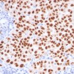
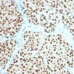
.jpg "SOX10 IHC Human Melanoma (SOX10 1074)")
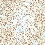
.jpg "SOX10 IHC Mouse (SOX10 1074)")
%20Gel.jpg "Anti-SOX10(SOX10 1074) Gel")
.jpg "SOX10 westerns (recombinant SOX10 and A375 lysate)")

 PDF Datasheet
Human Anti-SOX10 Antibody Product Attributes
SOX10 Previously Observed Antibody Staining Patterns
Observed Subcellular, Organelle Specific Staining Data:
Anti-SOX10 antibody staining is expected to be primarily localized to the nucleoplasm. There is variability in either the signal strength or the localization of signal in nucleoplasm from cell to cell.
Observed Antibody Staining Data By Tissue Type:
Variations in SOX10 antibody staining intensity in immunohistochemistry on tissue sections are present across different anatomical locations. An intense signal was observed in glial cells in the caudate nucleus and hippocampus, glandular cells in the salivary gland and melanocytes in skin. More moderate antibody staining intensity was present in glial cells in the caudate nucleus and hippocampus, glandular cells in the salivary gland and melanocytes in skin. Low, but measureable presence of SOX10 could be seen inglandular cells in the breast, cells in the granular layer in cerebellum, keratinocytes in skin, Langerhans in skin, epidermal cells in the skin, peripheral nerve in mesenchymal tissue and chondrocytes in mesenchymal tissue. We were unable to detect SOX10 in other tissues. Disease states, inflammation, and other physiological changes can have a substantial impact on antibody staining patterns. These measurements were all taken in tissues deemed normal or from patients without known disease.
Observed Antibody Staining Data By Tissue Disease Status:
Tissues from cancer patients, for instance, have their own distinct pattern of SOX10 expression as measured by anti-SOX10 antibody immunohistochemical staining. The average level of expression by tumor is summarized in the table below. The variability row represents patient to patient variability in IHC staining.
| Sample Type | breast cancer | carcinoid | cervical cancer | colorectal cancer | endometrial cancer | glioma | head and neck cancer | liver cancer | lung cancer | lymphoma | melanoma | ovarian cancer | pancreatic cancer | prostate cancer | renal cancer | skin cancer | stomach cancer | testicular cancer | thyroid cancer | urothelial cancer |
|---|---|---|---|---|---|---|---|---|---|---|---|---|---|---|---|---|---|---|---|---|
| Signal Intensity | – | – | – | – | – | + | – | – | – | – | + | – | – | – | – | – | – | – | – | – |
| SOX10 Variability | + | + | + | + | + | ++ | + | + | + | + | ++ | + | + | + | + | + | + | + | + | + |
| SOX10 General Information | |
|---|---|
| Alternate Names | |
| Transcription factor SOX-10, SOX10 | |
| Molecular Weight | |
| 49-58kDa | |
| Chromosomal Location | |
| 22q13.1 | |
| Curated Database and Bioinformatic Data | |
| Gene Symbol | SOX10 |
| Entrez Gene ID | 6663 |
| Ensemble Gene ID | ENSG00000100146 |
| RefSeq Protein Accession(s) | NP_008872 |
| RefSeq mRNA Accession(s) | NM_006941 |
| RefSeq Genomic Accession(s) | NG_007948, NC_000022, NC_018933 |
| UniProt ID(s) | P56693, A0A024R1N6 |
| UniGene ID(s) | P56693, A0A024R1N6 |
| HGNC ID(s) | 11190 |
| Cosmic ID(s) | SOX10 |
| KEGG Gene ID(s) | hsa:6663 |
| PharmGKB ID(s) | PA36027 |
| General Description of SOX10. | |
| Recognizes a protein of ~55kDa, identified as SOX10. This MAb is highly specific, does not cross-react with other members of the SOX-family. SOX genes comprise a family of genes that are related to the mammalian sex-determining gene SRY. These genes similarly contain sequences that encode for the HMG-box domain, which is responsible for the sequence-specific DNA-binding activity. SOX-10 is a sensitive marker of melanoma, including conventional, spindled,, desmoplastic subtypes. It is expressed by metastatic melanomas, nodal capsular nevus in sentinel lymph nodes, but not by other lymph node components such as dendritic cells, which usually express S100 protein. Commonly used melanoma markers, such as anti-HMB-45, anti-Melan-A, are poorly expressed in desmoplastic melanomas while SOX-10 is moderately to strongly expressed in desmoplastic melanomas. SOX-10 is considered as a very reliable marker for recognizing residual desmoplastic melanomas. In normal tissues, it is expressed in Schwann cells, melanocytes,, myoepithelial cells of salivary, bronchial, mammary glands. SOX-10 expression is also observed in mast cells. | |
There are no reviews yet.