
![Analysis of Mass Spec data (dashed-line) of fractions stained with SUMO-1 MS-QAVA™ monoclonal antibody [Clone: SUMO1/1188] (solid-line), reveals that less than 9.7% of signal is attributable to non-specific binding of anti-SUMO-1 [Clone SUMO1/1188] to targets other than SUMO1 protein. Even frequently cited antibodies have much greater non-specific interactions, averaging over 30%. Data in image is from analysis in A431, RT4 and MCF7 cells.](https://cdn-enquirebio.pressidium.com/wp-content/uploads/2017/10/enQuire-Bio-7341-MSM2-P1-anti-SUMO-1-antibody.png)
 PDF Datasheet
Human and Rat Anti-SUMO-1 Antibody Product Attributes
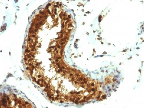
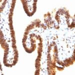
SUMO-1 Previously Observed Antibody Staining Patterns
Observed Subcellular, Organelle Specific Staining Data:
Variations in SUMO-1 antibody staining intensity in immunohistochemistry on tissue sections are present across different anatomical locations. An intense signal was observed in glandular cells in the adrenal gland, hematopoietic cells in the bone marrow, adipocytes in breast, respiratory epithelial cells in the bronchus, glandular cells in the duodenum, endometrium, epididymis, fallopian tube and gallbladder, macrophages in lung, pneumocytes in lung, respiratory epithelial cells in the nasopharynx, glandular cells in the parathyroid gland, decidual cells in the placenta, trophoblastic cells in the placenta, glandular cells in the rectum, myocytes in skeletal muscle, fibroblasts in skin, keratinocytes in skin, Langerhans in skin, melanocytes in skin, glandular cells in the small intestine, adipocytes in mesenchymal tissue, fibroblasts in mesenchymal tissue, cells in the seminiferous ducts in testis, germinal center cells in the tonsil, non-germinal center cells in the tonsil and urothelial cells in the urinary bladder. More moderate antibody staining intensity was present in glandular cells in the adrenal gland, hematopoietic cells in the bone marrow, adipocytes in breast, respiratory epithelial cells in the bronchus, glandular cells in the duodenum, endometrium, epididymis, fallopian tube and gallbladder, macrophages in lung, pneumocytes in lung, respiratory epithelial cells in the nasopharynx, glandular cells in the parathyroid gland, decidual cells in the placenta, trophoblastic cells in the placenta, glandular cells in the rectum, myocytes in skeletal muscle, fibroblasts in skin, keratinocytes in skin, Langerhans in skin, melanocytes in skin, glandular cells in the small intestine, adipocytes in mesenchymal tissue, fibroblasts in mesenchymal tissue, cells in the seminiferous ducts in testis, germinal center cells in the tonsil, non-germinal center cells in the tonsil and urothelial cells in the urinary bladder. Low, but measureable presence of SUMO-1 could be seen inglandular cells in the appendix, lymphoid tissue in appendix, glial cells in the caudate nucleus and cerebral cortex, bile duct cells in the liver and Leydig cells in the testis. We were unable to detect SUMO-1 in other tissues. Disease states, inflammation, and other physiological changes can have a substantial impact on antibody staining patterns. These measurements were all taken in tissues deemed normal or from patients without known disease.
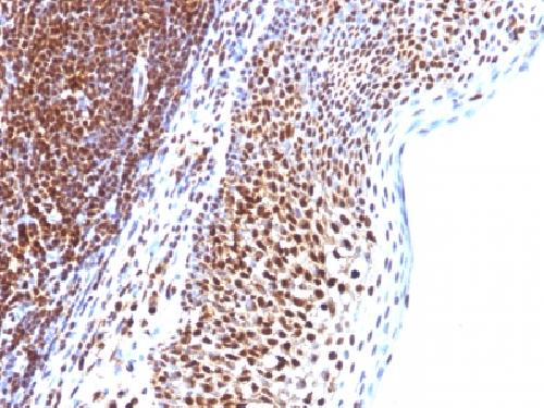
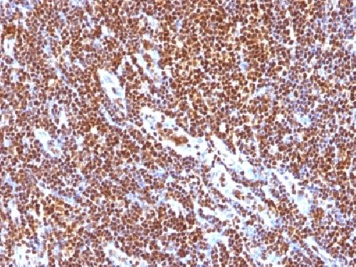
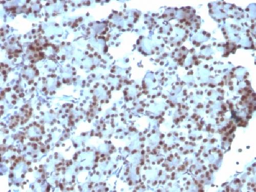
Observed Antibody Staining Data By Tissue Type:
Tissues from cancer patients, for instance, have their own distinct pattern of SUMO-1 expression as measured by anti-SUMO-1 antibody immunohistochemical staining. The average level of expression by tumor is summarized in the table below. The variability row represents patient to patient variability in IHC staining.
| Sample Type | breast cancer | carcinoid | cervical cancer | colorectal cancer | endometrial cancer | glioma | head and neck cancer | liver cancer | lung cancer | lymphoma | melanoma | ovarian cancer | pancreatic cancer | prostate cancer | renal cancer | skin cancer | stomach cancer | testicular cancer | thyroid cancer | urothelial cancer |
|---|---|---|---|---|---|---|---|---|---|---|---|---|---|---|---|---|---|---|---|---|
| Signal Intensity | ++ | + | ++ | ++ | + | ++ | ++ | ++ | + | ++ | ++ | +++ | ++ | ++ | + | ++ | ++ | +++ | ++ | ++ |
| SUMO1 Variability | ++ | ++ | ++ | ++ | +++ | ++ | ++ | +++ | ++ | ++ | +++ | ++ | ++ | ++ | +++ | ++ | ++ | ++ | ++ | +++ |
| SUMO-1 General Information | |
|---|---|
| Alternate Names | |
| Small ubiquitin-related modifier 1, SUMO1, SUMO-1 | |
| Molecular Weight | |
| 11.5kDa (Monomer); 90kDa (Heteromer) | |
| Chromosomal Location | |
| 2q33.1 | |
| Curated Database and Bioinformatic Data | |
| Gene Symbol | SUMO1 |
| Entrez Gene ID | 7341 |
| Ensemble Gene ID | ENSG00000116030 |
| RefSeq Protein Accession(s) | NP_003343, NP_001005781, NP_001005782 |
| RefSeq mRNA Accession(s) | NM_003352, NM_001005782 NM_001005781 |
| RefSeq Genomic Accession(s) | NG_011679, NC_000002, NC_018913 |
| UniProt ID(s) | P55856, A0A024R3Z2, Q93068, P63165 |
| UniGene ID(s) | P55856, A0A024R3Z2, Q93068, P63165 |
| HGNC ID(s) | 12502 |
| Cosmic ID(s) | SUMO1 |
| KEGG Gene ID(s) | hsa:7341 |
| PharmGKB ID(s) | PA37149 |
| General Description of SUMO-1. | |
| This MAb is specific to SUMO-1, shows no cross-reaction with either SUMO-2 or SUMO-3. The small ubiquitin-related modifier (SUMO) proteins, which include SUMO-1, SUMO-2, SUMO-3, belong to the ubiquitin-like protein family. Like ubiquitin, the SUMO proteins are synthesized as precursor proteins that undergo processing before conjugation to target proteins. Also, both utilize the E1, E2,, E3 cascade enzymes for conjugation. However, SUMO, ubiquitin differ with respect to targeting. Ubiquitination predominantly targets proteins for degradation, whereas sumoylation targets proteins to a variety of cellular processing, including nuclear transport, transcriptional regulation, apoptosis, protein stability. The unconjugated SUMO-1 protein localizes to the nuclear membrane. | |

Reviews
There are no reviews yet.