
 PDF Datasheet
Human, Rabbit, and Zebrafish Anti-TGF-alpha Antibody Product Attributes
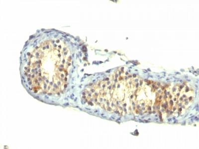
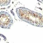
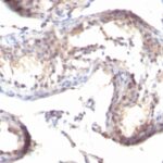
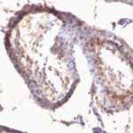
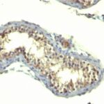
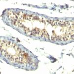
TGF-alpha Previously Observed Antibody Staining Patterns
Observed Subcellular, Organelle Specific Staining Data:
Anti-TGFA antibody staining is expected to be primarily localized to the vesicles. There is variability in either the signal strength or the localization of signal in vesicles from cell to cell.
Observed Antibody Staining Data By Tissue Type:
Variations in TGF-alpha antibody staining intensity in immunohistochemistry on tissue sections are present across different anatomical locations. Low, but measureable presence of TGF-alpha could be seen inlymphoid tissue in appendix, hematopoietic cells in the bone marrow, glial cells in the caudate nucleus, cells in the granular layer in cerebellum, endothelial cells in the colon, glandular cells in the endometrium, neuronal cells in the hippocampus, pneumocytes in lung, germinal center cells in the lymph node, squamous epithelial cells in the oral mucosa, epidermal cells in the skin, smooth muscle cells in the smooth muscle, fibroblasts in mesenchymal tissue, peripheral nerve in mesenchymal tissue and non-germinal center cells in the tonsil. We were unable to detect TGF-alpha in other tissues. Disease states, inflammation, and other physiological changes can have a substantial impact on antibody staining patterns. These measurements were all taken in tissues deemed normal or from patients without known disease.
Observed Antibody Staining Data By Tissue Disease Status:
Tissues from cancer patients, for instance, have their own distinct pattern of TGF-alpha expression as measured by anti-TGF-alpha antibody immunohistochemical staining. The average level of expression by tumor is summarized in the table below. The variability row represents patient to patient variability in IHC staining.
| Sample Type | breast cancer | carcinoid | cervical cancer | colorectal cancer | endometrial cancer | glioma | head and neck cancer | liver cancer | lung cancer | lymphoma | melanoma | ovarian cancer | pancreatic cancer | prostate cancer | renal cancer | skin cancer | stomach cancer | testicular cancer | thyroid cancer | urothelial cancer |
|---|---|---|---|---|---|---|---|---|---|---|---|---|---|---|---|---|---|---|---|---|
| Signal Intensity | ++ | – | + | + | ++ | ++ | – | ++ | ++ | + | ++ | ++ | ++ | ++ | ++ | – | ++ | – | ++ | + |
| TGFA Variability | ++ | ++ | +++ | ++ | ++ | ++ | ++ | ++ | ++ | ++ | ++ | ++ | ++ | + | ++ | ++ | ++ | ++ | ++ | ++ |
| TGF-alpha General Information | |
|---|---|
| Alternate Names | |
| Transforming growth factor alpha, TGF-?, TGFa, TGF alpha | |
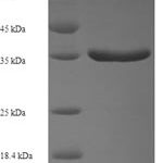
| Molecular Weight | |
| ~6kDa | |
| Chromosomal Location | |
| 2p13.3 | |
| Curated Database and Bioinformatic Data | |
| Gene Symbol | TGFA |
| Entrez Gene ID | 7039 |
| Ensemble Gene ID | ENSG00000163235 |
| RefSeq Protein Accession(s) | NP_001295088, NP_001093161, NP_003227, NP_001295087 |
| RefSeq mRNA Accession(s) | NM_001308158, NM_003236, NM_001099691 NM_001308159 |
| RefSeq Genomic Accession(s) | NG_029975, NC_018913, NC_000002 |
| UniProt ID(s) | F8VNR3, P01135, E7EPT6 |
| UniGene ID(s) | F8VNR3, P01135, E7EPT6 |
| HGNC ID(s) | 11765 |
| Cosmic ID(s) | TGFA |
| KEGG Gene ID(s) | hsa:7039 |
| PharmGKB ID(s) | PA36480 |
| General Description of TGF-alpha. | |
| This antibody reacts with the TGF alpha, shows no cross-reaction with EGF, the neuropeptide synenkephalin. The staining with this MAb is completely blocked by the peptide used for raising this antibody. TGF? (aa50) is a growth factor with 33% homology to EGF, binds to EGFR, activates tyrosine phosphorylation of the receptor,, stimulates cell proliferation. It plays a role in tumor initiation by inducing the reversible transformed phenotype. | |
Reviews
There are no reviews yet.