

 PDF Datasheet
Human, Mouse, and Rat Anti-Thymidine Phosphorylase / PD-ECGF Antibody Product Attributes
Thymidine Phosphorylase / PD-ECGF Previously Observed Antibody Staining Patterns
Observed Subcellular, Organelle Specific Staining Data:
Anti-TYMP antibody staining is expected to be primarily localized to the golgi apparatus and nuclear bodies.
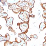
Observed Antibody Staining Data By Tissue Type:
Variations in Thymidine Phosphorylase / PD-ECGF antibody staining intensity in immunohistochemistry on tissue sections are present across different anatomical locations. Low, but measureable presence of Thymidine Phosphorylase / PD-ECGF could be seen inendothelial cells in the cerebral cortex, glial cells in the cerebral cortex, cells in the endometrial stroma in endometrium, glandular cells in the endometrium, cells in the tubules in kidney, glandular cells in the prostate and seminal vesicle, keratinocytes in skin, melanocytes in skin, non-germinal center cells in the tonsil and urothelial cells in the urinary bladder. We were unable to detect Thymidine Phosphorylase / PD-ECGF in other tissues. Disease states, inflammation, and other physiological changes can have a substantial impact on antibody staining patterns. These measurements were all taken in tissues deemed normal or from patients without known disease.
Observed Antibody Staining Data By Tissue Disease Status:
Tissues from cancer patients, for instance, have their own distinct pattern of Thymidine Phosphorylase / PD-ECGF expression as measured by anti-Thymidine Phosphorylase / PD-ECGF antibody immunohistochemical staining. The average level of expression by tumor is summarized in the table below. The variability row represents patient to patient variability in IHC staining.
| Sample Type | breast cancer | carcinoid | cervical cancer | colorectal cancer | endometrial cancer | glioma | head and neck cancer | liver cancer | lung cancer | lymphoma | melanoma | ovarian cancer | pancreatic cancer | prostate cancer | renal cancer | skin cancer | stomach cancer | testicular cancer | thyroid cancer | urothelial cancer |
|---|---|---|---|---|---|---|---|---|---|---|---|---|---|---|---|---|---|---|---|---|
| Signal Intensity | + | – | ++ | + | – | + | ++ | + | – | + | + | + | – | – | – | – | – | + | + | + |
| TYMP Variability | +++ | + | ++ | +++ | ++ | ++ | ++ | ++ | ++ | ++ | +++ | ++ | ++ | ++ | ++ | ++ | ++ | ++ | ++ | ++ |
| Thymidine Phosphorylase / PD-ECGF General Information | |
|---|---|
| Alternate Names | |
| Thymidine Phosphorylase, TYMP gene, ECGF1, Platelet-derived endothelial cell growth factor | |
| Molecular Weight | |
| 55kDa | |
| Chromosomal Location | |
| 22q13.33 | |
| Curated Database and Bioinformatic Data | |
| Gene Symbol | TYMP |
| Entrez Gene ID | 1890 |
| Ensemble Gene ID | ENSG00000025708 |
| RefSeq Protein Accession(s) | NP_001944, NP_001107228, NP_001244918, NP_001244917, NP_001107227 |
| RefSeq mRNA Accession(s) | NM_001257988, NM_001113756, NM_001257989 NM_001113755, NM_001953 |
| RefSeq Genomic Accession(s) | NG_011860, NC_018933, NC_000022, NG_016235 |
| UniProt ID(s) | P19971, E5KRG5, B2RBL3 |
| UniGene ID(s) | P19971, E5KRG5, B2RBL3 |
| HGNC ID(s) | 3148 |
| Cosmic ID(s) | TYMP |
| KEGG Gene ID(s) | hsa:1890 |
| PharmGKB ID(s) | PA162407502 |
| General Description of Thymidine Phosphorylase / PD-ECGF. | |
| Recognizes a protein (amino acid 482) of 55kDa (in vivo 110kDa homodimer), identified as platelet-derived endothelial growth factor (PD-ECGF), same as thymidine phosphorylase (TP) or gliostatin. In the presence of inorganic orthophosphate, it catalyzes the reversible phospholytic cleavage of thymidine and deoxyuridine to their corresponding bases and 2-deoxyribose-1-phosphate. It is both chemotactic and mitogenic for endothelial cells and a non-heparin binding angiogenic factor present in platelets. Its enzymatic activity is crucial for angiogenic activity (metabolite is angiogenic). Higher levels of serum TP/PD-ECGF are observed in cancer patients. It is also involved in transformation of fluoropyrimidines, cytotoxic agents used in the treatment of a variety of malignancies, into active cytotoxic metabolites (e.g. 5 -deoxy-5-fluorouridine to 5-FU). High intra-cellular levels of TP/PD-ECGF are associated with increased chemosensitivity to such antimetabolites. | |
There are no reviews yet.