
 PDF Datasheet
Human Anti-Topoisomerase I, Mitochondrial Antibody Product Attributes
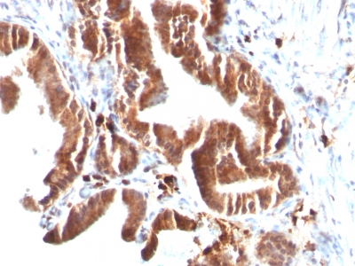
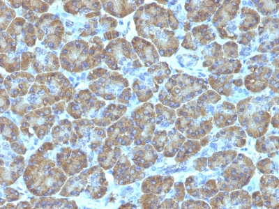
Topoisomerase I, Mitochondrial Previously Observed Antibody Staining Patterns
Observed Subcellular, Organelle Specific Staining Data:
Anti-TOP1MT antibody staining is expected to be primarily localized to the mitochondria.
Observed Antibody Staining Data By Tissue Type:
Variations in Topoisomerase I, Mitochondrial antibody staining intensity in immunohistochemistry on tissue sections are present across different anatomical locations. An intense signal was observed in glandular cells in the appendix, hematopoietic cells in the bone marrow, glandular cells in the breast, myoepithelial cells in the breast, neuronal cells in the caudate nucleus, Purkinje cells in the cerebellum, neuronal cells in the cerebral cortex, squamous epithelial cells in the cervix, uterine, glandular cells in the colon, duodenum, endometrium, epididymis, fallopian tube and gallbladder, myocytes in heart muscle, neuronal cells in the hippocampus, cells in the tubules in kidney, hepatocytes in liver, macrophages in lung, pneumocytes in lung, respiratory epithelial cells in the nasopharynx, exocrine glandular cells in the pancreas, glandular cells in the parathyroid gland, decidual cells in the placenta, trophoblastic cells in the placenta, glandular cells in the prostate, small intestine and stomach, cells in the seminiferous ducts in testis, glandular cells in the thyroid gland, germinal center cells in the tonsil, non-germinal center cells in the tonsil and urothelial cells in the urinary bladder. More moderate antibody staining intensity was present in glandular cells in the appendix, hematopoietic cells in the bone marrow, glandular cells in the breast, myoepithelial cells in the breast, neuronal cells in the caudate nucleus, Purkinje cells in the cerebellum, neuronal cells in the cerebral cortex, squamous epithelial cells in the cervix, uterine, glandular cells in the colon, duodenum, endometrium, epididymis, fallopian tube and gallbladder, myocytes in heart muscle, neuronal cells in the hippocampus, cells in the tubules in kidney, hepatocytes in liver, macrophages in lung, pneumocytes in lung, respiratory epithelial cells in the nasopharynx, exocrine glandular cells in the pancreas, glandular cells in the parathyroid gland, decidual cells in the placenta, trophoblastic cells in the placenta, glandular cells in the prostate, small intestine and stomach, cells in the seminiferous ducts in testis, glandular cells in the thyroid gland, germinal center cells in the tonsil, non-germinal center cells in the tonsil and urothelial cells in the urinary bladder. Low, but measureable presence of Topoisomerase I, Mitochondrial could be seen in cells in the endometrial stroma in endometrium, glial cells in the hippocampus, germinal center cells in the lymph node, Langerhans in skin, melanocytes in skin, smooth muscle cells in the smooth muscle, adipocytes in mesenchymal tissue and cells in the white pulp in spleen. We were unable to detect Topoisomerase I, Mitochondrial in other tissues. Disease states, inflammation, and other physiological changes can have a substantial impact on antibody staining patterns. These measurements were all taken in tissues deemed normal or from patients without known disease.
Observed Antibody Staining Data By Tissue Disease Status:
Tissues from cancer patients, for instance, have their own distinct pattern of Topoisomerase I, Mitochondrial expression as measured by anti-Topoisomerase I, Mitochondrial antibody immunohistochemical staining. The average level of expression by tumor is summarized in the table below. The variability row represents patient to patient variability in IHC staining.
| Sample Type | breast cancer | carcinoid | cervical cancer | colorectal cancer | endometrial cancer | glioma | head and neck cancer | liver cancer | lung cancer | lymphoma | melanoma | ovarian cancer | pancreatic cancer | prostate cancer | renal cancer | skin cancer | stomach cancer | testicular cancer | thyroid cancer | urothelial cancer |
|---|---|---|---|---|---|---|---|---|---|---|---|---|---|---|---|---|---|---|---|---|
| Signal Intensity | ++ | + | ++ | +++ | +++ | ++ | ++ | +++ | ++ | ++ | ++ | ++ | ++ | ++ | + | ++ | ++ | ++ | ++ | ++ |
| TOP1MT Variability | ++ | ++ | ++ | ++ | ++ | ++ | +++ | ++ | ++ | + | ++ | ++ | + | + | ++ | ++ | ++ | + | ++ | ++ |
| Topoisomerase I, Mitochondrial General Information | |
|---|---|
| Alternate Names | |
| Aspartate aminotransferase, Cytoplasmic Aspartate Aminotransferase | |
| Molecular Weight | |
| 70kDa | |
| Chromosomal Location | |
| 8q24.3 | |
| Curated Database and Bioinformatic Data | |
| Gene Symbol | TOP1MT |
| Entrez Gene ID | 116447 |
| Ensemble Gene ID | ENSG00000184428 |
| RefSeq Protein Accession(s) | XP_005250838, NP_443195, NP_001245375, NP_001245376 |
| RefSeq mRNA Accession(s) | XM_005250781, NM_052963, NM_001258446, NM_001258447 |
| RefSeq Genomic Accession(s) | NC_000008, NC_018919 |
| UniProt ID(s) | Q969P6, E5KMK7, B4DYD2 |
| UniGene ID(s) | Q969P6, E5KMK7, B4DYD2 |
| HGNC ID(s) | 29787 |
| Cosmic ID(s) | TOP1MT |
| KEGG Gene ID(s) | hsa:116447 |
| PharmGKB ID(s) | PA134922772 |
| General Description of Topoisomerase I, Mitochondrial. | |
| DNA topoisomerases are nuclear enzymes that regulate the topological structure of DNA in eukaryotic cells by transiently breaking, rejoining DNA strands. Due to their roles in DNA replication, recombination,, transcription, DNA topoisomerases have been identified as targets of numerous anticancer drugs. Mitochondrial Topo I (DNA topoisomerase I, mitochondrial) is a 601 amino acid protein that primarily acts to relieve DNA strain that may occur during duplication of mitochondrial DNA. As a type IB topoisomerase, mitochondrial Topo I requires a divalent metal, either, calcium or magnesium, as well as an alkaline pH for optimal activity. | |
Reviews
There are no reviews yet.