
 PDF Datasheet
Human, Monkey, Rabbit, Mouse, and Rat Anti-Transglutaminase II Antibody Product Attributes
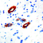
Transglutaminase II Previously Observed Antibody Staining Patterns
Observed Subcellular, Organelle Specific Staining Data:
Anti-TGM2 antibody staining is expected to be primarily localized to the cytosol and plasma membrane. There is variability in either the signal strength or the localization of signal in cytosol and plasma membrane from cell to cell.
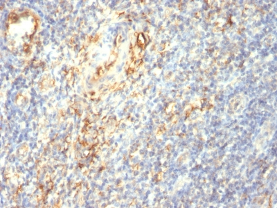
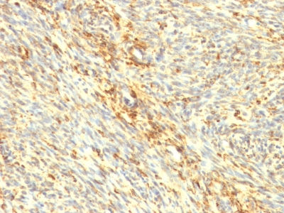
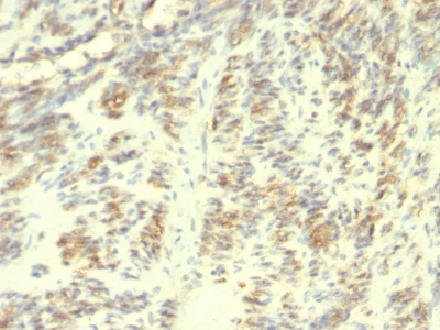
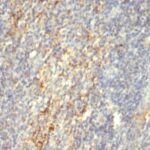
Observed Antibody Staining Data By Tissue Type:
Variations in Transglutaminase II antibody staining intensity in immunohistochemistry on tissue sections are present across different anatomical locations. An intense signal was observed in decidual cells in the placenta and trophoblastic cells in the placenta. More moderate antibody staining intensity was present in decidual cells in the placenta and trophoblastic cells in the placenta. Low, but measureable presence of Transglutaminase II could be seen inlymphoid tissue in appendix, endothelial cells in the cerebral cortex, glandular cells in the cervix, uterine, squamous epithelial cells in the cervix, uterine, melanocytes in skin, adipocytes in mesenchymal tissue, glandular cells in the stomach, Leydig cells in the testis and urothelial cells in the urinary bladder. We were unable to detect Transglutaminase II in other tissues. Disease states, inflammation, and other physiological changes can have a substantial impact on antibody staining patterns. These measurements were all taken in tissues deemed normal or from patients without known disease.
| Transglutaminase II General Information | |
|---|---|
| Alternate Names | |
| Tissue transglutaminase, tTG, TG2 | |
| Molecular Weight | |
| 77-85kDa | |
| Chromosomal Location | |
| 20q12 | |
| Curated Database and Bioinformatic Data | |
| Gene Symbol | TGM2 |
| Entrez Gene ID | 7052 |
| Ensemble Gene ID | ENSG00000198959 |
| RefSeq Protein Accession(s) | NP_001310247, NP_001310246, NP_945189, NP_001310245, XP_011527330, NP_004604 |
| RefSeq mRNA Accession(s) | NM_198951, NM_001323316, NM_001323317, NM_004613, XM_011529028, NM_001323318 |
| RefSeq Genomic Accession(s) | NC_000020, NC_018931 |
| UniProt ID(s) | B4DTN7, P21980, B4DIT7, V9HWG3 |
| UniGene ID(s) | B4DTN7, P21980, B4DIT7, V9HWG3 |
| HGNC ID(s) | 11778 |
| Cosmic ID(s) | TGM2 |
| KEGG Gene ID(s) | hsa:7052 |
| PharmGKB ID(s) | PA36491 |
| General Description of Transglutaminase II. | |
| Recognizes a 77-85kDa protein, identified as cellular or tissue transglutaminase II (TGase II). Transglutaminases are enzymes that catalyze the crosslinking of proteins by epsilon-gamma glutamyl lysine isopeptide bonds. While the primary structure of transglutaminases is not conserved, they all have the same amino acid sequence at their active sites, their activity is calcium-dependent. The protein encoded by this gene acts as a monomer, is induced by retinoic acid,, appears to be involved in apoptosis. Finally, the encoded protein is the autoantigen implicated in celiac disease. The identification of transglutaminase as the main antigen of endomysium antibodies allows a new diagnostic approach to celiac disease (CD), a genetic, immunologically mediated small bowel enteropathy that causes malabsorption. TGase II is implicated in programmed cell death, signal transduction, drug-resistance, cell growth, endocytosis, insulin secretion, cell adhesion, cataract formation,, wound healing. | |
Reviews
There are no reviews yet.