
 PDF Datasheet
Human, Mouse, and Rat Anti-TTF-1 / NKX2.1 Antibody Product Attributes
TTF-1 / NKX2.1 Previously Observed Antibody Staining Patterns
Observed Antibody Staining Data By Tissue Type:
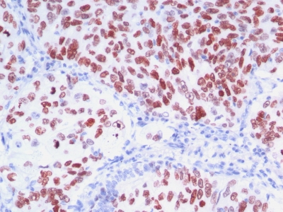
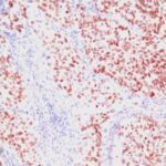
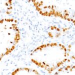
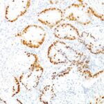
Variations in TTF-1 / NKX2.1 antibody staining intensity in immunohistochemistry on tissue sections are present across different anatomical locations. An intense signal was observed in glandular cells in the thyroid gland. More moderate antibody staining intensity was present in glandular cells in the thyroid gland. Low, but measureable presence of TTF-1 / NKX2.1 could be seen inrespiratory epithelial cells in the bronchus, macrophages in lung and respiratory epithelial cells in the nasopharynx. We were unable to detect TTF-1 / NKX2.1 in other tissues. Disease states, inflammation, and other physiological changes can have a substantial impact on antibody staining patterns. These measurements were all taken in tissues deemed normal or from patients without known disease.
Observed Antibody Staining Data By Tissue Disease Status:
Tissues from cancer patients, for instance, have their own distinct pattern of TTF-1 / NKX2.1 expression as measured by anti-TTF-1 / NKX2.1 antibody immunohistochemical staining. The average level of expression by tumor is summarized in the table below. The variability row represents patient to patient variability in IHC staining.
| Sample Type | breast cancer | carcinoid | cervical cancer | colorectal cancer | endometrial cancer | glioma | head and neck cancer | liver cancer | lung cancer | lymphoma | melanoma | ovarian cancer | pancreatic cancer | prostate cancer | renal cancer | skin cancer | stomach cancer | testicular cancer | thyroid cancer | urothelial cancer |
|---|---|---|---|---|---|---|---|---|---|---|---|---|---|---|---|---|---|---|---|---|
| Signal Intensity | – | – | – | – | – | – | – | – | ++ | – | – | – | – | – | – | – | – | – | ++ | – |
| NKX2-1 Variability | + | + | + | + | + | + | + | ++ | ++ | + | + | + | + | + | + | + | + | + | ++ | + |
| TTF-1 / NKX2.1 General Information | |
|---|---|
| Alternate Names | |
| NK2 homeobox 1, NKX2-1, thyroid transcription factor 1, TTF-1 | |
| Molecular Weight | |
| 40kDa | |
| Chromosomal Location | |
| 14q13.3 | |
| Curated Database and Bioinformatic Data | |
| Gene Symbol | NKX2-1 |
| Entrez Gene ID | 7080 |
| Ensemble Gene ID | ENSG00000136352 |
| RefSeq Protein Accession(s) | NP_001073136, NP_003308 |
| RefSeq mRNA Accession(s) | NM_003317 NM_001079668 |
| RefSeq Genomic Accession(s) | NC_018925, NG_013365, NC_000014 |
| UniProt ID(s) | P43699 |
| UniGene ID(s) | P43699 |
| HGNC ID(s) | 11825 |
| Cosmic ID(s) | NKX2-1 |
| KEGG Gene ID(s) | hsa:7080 |
| PharmGKB ID(s) | PA36531 |
| General Description of TTF-1 / NKX2.1. | |
| Recognizes a protein of 40kDa, identified as Thyroid transcription factor-1 (TTF-1). TTF-1 is a member of the NKx2 family of homeodomain transcription factors. It is expressed in epithelial cells of the thyroid gland, the lung. Nuclei from liver, stomach, pancreas, small intestine, colon, kidney, breast, skin, testes, pituitary, prostate,, adrenal glands are unreactive. Anti-TTF-1 is useful in differentiating primary adenocarcinoma of the lung from metastatic carcinomas originating in the breast, mediastinal germ cell tumors,, malignant mesothelioma. It can also be used to differentiate small cell lung carcinoma from lymphoid infiltrates.Loss of TTF-1 expression in non-small cell lung carcinoma has been associated with aggressive behavior of such neoplasms. TTF-1 reactivity is also seen in thyroid malignancies. | |
There are no reviews yet.