

 PDF Datasheet
Human and Mouse Anti-Tyrosinase-Related Protein-1 Antibody Product Attributes
Tyrosinase-Related Protein-1 Previously Observed Antibody Staining Patterns
Observed Subcellular, Organelle Specific Staining Data:
Anti-TYRP1 antibody staining is expected to be primarily localized to the vesicles.
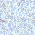
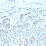
Observed Antibody Staining Data By Tissue Type:
Variations in Tyrosinase-Related Protein-1 antibody staining intensity in immunohistochemistry on tissue sections are present across different anatomical locations. An intense signal was observed in melanocytes in skin. More moderate antibody staining intensity was present in melanocytes in skin. Low, but measureable presence of Tyrosinase-Related Protein-1 could be seen in. We were unable to detect Tyrosinase-Related Protein-1 in other tissues. Disease states, inflammation, and other physiological changes can have a substantial impact on antibody staining patterns. These measurements were all taken in tissues deemed normal or from patients without known disease.
Observed Antibody Staining Data By Tissue Disease Status:
Tissues from cancer patients, for instance, have their own distinct pattern of Tyrosinase-Related Protein-1 expression as measured by anti-Tyrosinase-Related Protein-1 antibody immunohistochemical staining. The average level of expression by tumor is summarized in the table below. The variability row represents patient to patient variability in IHC staining.
| Sample Type | breast cancer | carcinoid | cervical cancer | colorectal cancer | endometrial cancer | glioma | head and neck cancer | liver cancer | lung cancer | lymphoma | melanoma | ovarian cancer | pancreatic cancer | prostate cancer | renal cancer | skin cancer | stomach cancer | testicular cancer | thyroid cancer | urothelial cancer |
|---|---|---|---|---|---|---|---|---|---|---|---|---|---|---|---|---|---|---|---|---|
| Signal Intensity | – | – | – | – | – | – | – | – | – | – | ++ | – | – | – | – | – | – | – | – | – |
| TYRP1 Variability | + | + | + | ++ | + | + | ++ | + | + | + | ++ | + | + | + | + | ++ | + | ++ | + | + |
| Tyrosinase-Related Protein-1 General Information | |
|---|---|
| Alternate Names | |
| Tyrosinase-related protein 1, TYRP1 | |
| Molecular Weight | |
| 75kDa | |
| Chromosomal Location | |
| 9p23 | |
| Curated Database and Bioinformatic Data | |
| Gene Symbol | TYRP1 |
| Entrez Gene ID | 7306 |
| Ensemble Gene ID | ENSG00000107165 |
| RefSeq Protein Accession(s) | NP_000541 |
| RefSeq mRNA Accession(s) | NM_000550, XR_001746372, |
| RefSeq Genomic Accession(s) | NG_011705, NC_018920, NC_000009 |
| UniProt ID(s) | P17643 |
| UniGene ID(s) | P17643 |
| HGNC ID(s) | 12450 |
| Cosmic ID(s) | TYRP1 |
| KEGG Gene ID(s) | hsa:7306 |
| PharmGKB ID(s) | PA37101 |
| General Description of Tyrosinase-Related Protein-1. | |
| It reacts with a 75kDa melanocyte-specific gene product, identified as Tyrosinase-related protein-1 (TRP-1). It is involved in melanin synthesis. TRP1 is present on the melanosomal membranes of melanoma, normal melanocytes and nevi. Recent evidence suggests that TRP-1 is involved in maintaining stability of tyrosinase protein and modulating its catalytic activity. TRP-1 is also involved in maintenance of melanosome ultrastructure and affects melanocyte proliferation and cell death. | |
There are no reviews yet.