
.jpg)
.jpg)
 PDF Datasheet
Human Anti-VEGI Antibody Product Attributes
VEGI Previously Observed Antibody Staining Patterns
Observed Subcellular, Organelle Specific Staining Data:
Anti-TNFSF15 antibody staining is expected to be primarily localized to the cytosol, nuclear membrane and nucleus.
Observed Antibody Staining Data By Tissue Type:
Variations in VEGI antibody staining intensity in immunohistochemistry on tissue sections are present across different anatomical locations. Low, but measureable presence of VEGI could be seen inglandular cells in the breast, myoepithelial cells in the breast, neuronal cells in the caudate nucleus, endothelial cells in the cerebral cortex, squamous epithelial cells in the cervix, uterine, endothelial cells in the colon, cells in the endometrial stroma in endometrium, cells in the glomeruli in kidney, bile duct cells in the liver, hepatocytes in liver, pneumocytes in lung, squamous epithelial cells in the oral mucosa, exocrine glandular cells in the pancreas, glandular cells in the salivary gland, keratinocytes in skin, melanocytes in skin, fibroblasts in mesenchymal tissue, germinal center cells in the tonsil, non-germinal center cells in the tonsil and squamous epithelial cells in the tonsil and vagina. We were unable to detect VEGI in other tissues. Disease states, inflammation, and other physiological changes can have a substantial impact on antibody staining patterns. These measurements were all taken in tissues deemed normal or from patients without known disease.
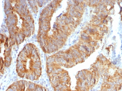
Observed Antibody Staining Data By Tissue Disease Status:
Tissues from cancer patients, for instance, have their own distinct pattern of VEGI expression as measured by anti-VEGI antibody immunohistochemical staining. The average level of expression by tumor is summarized in the table below. The variability row represents patient to patient variability in IHC staining.
| Sample Type | breast cancer | carcinoid | cervical cancer | colorectal cancer | endometrial cancer | glioma | head and neck cancer | liver cancer | lung cancer | lymphoma | melanoma | ovarian cancer | pancreatic cancer | prostate cancer | renal cancer | skin cancer | stomach cancer | testicular cancer | thyroid cancer | urothelial cancer |
|---|---|---|---|---|---|---|---|---|---|---|---|---|---|---|---|---|---|---|---|---|
| Signal Intensity | + | + | + | + | ++ | + | + | + | + | – | + | ++ | + | + | – | + | + | – | ++ | + |
| TNFSF15 Variability | + | ++ | ++ | ++ | ++ | +++ | ++ | ++ | ++ | ++ | ++ | ++ | ++ | +++ | ++ | ++ | ++ | ++ | ++ | ++ |
| VEGI General Information | |
|---|---|
| Alternate Names | |
| Vascular endothelial growth inhibitor, VEGI, TNF-like ligand 1A, TL1A, TNF superfamily member 15, TNFSF15 | |
| Molecular Weight | |
| 22kDa | |
| Chromosomal Location | |
| 9q32 | |
| Curated Database and Bioinformatic Data | |
| Gene Symbol | TNFSF15 |
| Entrez Gene ID | 9966 |
| Ensemble Gene ID | ENSG00000181634 |
| RefSeq Protein Accession(s) | NP_005109, NP_001191273 |
| RefSeq mRNA Accession(s) | NM_001204344, NM_005118 |
| RefSeq Genomic Accession(s) | NG_011488, NC_000009, NC_018920 |
| UniProt ID(s) | O95150, A0A0U5JA19 |
| UniGene ID(s) | O95150, A0A0U5JA19 |
| HGNC ID(s) | 11931 |
| Cosmic ID(s) | TNFSF15 |
| KEGG Gene ID(s) | hsa:9966 |
| PharmGKB ID(s) | PA36623 |
| General Description of VEGI. | |
| VEGI is an anti-angiogenic cytokine that belongs to tumor necrosis factor superfamily, member 15 (TNFSF15). This protein is abundantly expressed in endothelial cells, but is not expressed in either B or T cells. The expression of this protein is inducible by TNF and IL-1 alpha. This cytokine is a ligand for receptor TNFRSF25 and decoy receptor TNFRSF21/DR6. It can activate NF-kappaB and MAP kinases, and acts as an autocrine factor to induce apoptosis in endothelial cells. This cytokine is also found to inhibit endothelial cell proliferation, and thus may function as an angiogenesis inhibitor. Reduced expression of VEGI has been reported as a marker of poor prognosis in breast cancer. | |
Reviews
There are no reviews yet.