
 PDF Datasheet
Human, Cat, Cow, Pig, Rat, Rabbit, Mouse, and Sheep Anti-Vimentin Antibody Product Attributes
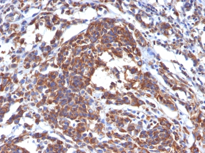
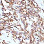
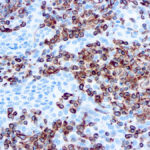
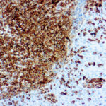
Vimentin Previously Observed Antibody Staining Patterns
Observed Subcellular, Organelle Specific Staining Data:
Anti-VIM antibody staining is expected to be primarily localized to the intermediate filaments.
Observed Antibody Staining Data By Tissue Type:
Variations in Vimentin antibody staining intensity in immunohistochemistry on tissue sections are present across different anatomical locations. An intense signal was observed in lymphoid tissue in appendix, hematopoietic cells in the bone marrow, adipocytes in breast, endothelial cells in the cerebral cortex and colon, glandular cells in the epididymis and fallopian tube, cells in the glomeruli in kidney, macrophages in lung, pneumocytes in lung, germinal center cells in the lymph node, non-germinal center cells in the lymph node, ovarian stroma cells in the ovary, glandular cells in the seminal vesicle, fibroblasts in skin, Langerhans in skin, melanocytes in skin, adipocytes in mesenchymal tissue, fibroblasts in mesenchymal tissue, peripheral nerve in mesenchymal tissue, cells in the red pulp in spleen, cells in the seminiferous ducts in testis, Leydig cells in the testis, glandular cells in the thyroid gland, germinal center cells in the tonsil and non-germinal center cells in the tonsil. More moderate antibody staining intensity was present in lymphoid tissue in appendix, hematopoietic cells in the bone marrow, adipocytes in breast, endothelial cells in the cerebral cortex and colon, glandular cells in the epididymis and fallopian tube, cells in the glomeruli in kidney, macrophages in lung, pneumocytes in lung, germinal center cells in the lymph node, non-germinal center cells in the lymph node, ovarian stroma cells in the ovary, glandular cells in the seminal vesicle, fibroblasts in skin, Langerhans in skin, melanocytes in skin, adipocytes in mesenchymal tissue, fibroblasts in mesenchymal tissue, peripheral nerve in mesenchymal tissue, cells in the red pulp in spleen, cells in the seminiferous ducts in testis, Leydig cells in the testis, glandular cells in the thyroid gland, germinal center cells in the tonsil and non-germinal center cells in the tonsil. Low, but measureable presence of Vimentin could be seen inglandular cells in the adrenal gland, respiratory epithelial cells in the bronchus, glial cells in the cerebral cortex, cells in the endometrial stroma in endometrium, glial cells in the hippocampus, bile duct cells in the liver, respiratory epithelial cells in the nasopharynx, follicle cells in the ovary, decidual cells in the placenta and squamous epithelial cells in the vagina. We were unable to detect Vimentin in other tissues. Disease states, inflammation, and other physiological changes can have a substantial impact on antibody staining patterns. These measurements were all taken in tissues deemed normal or from patients without known disease.
Observed Antibody Staining Data By Tissue Disease Status:
Tissues from cancer patients, for instance, have their own distinct pattern of Vimentin expression as measured by anti-Vimentin antibody immunohistochemical staining. The average level of expression by tumor is summarized in the table below. The variability row represents patient to patient variability in IHC staining.
| Sample Type | breast cancer | carcinoid | cervical cancer | colorectal cancer | endometrial cancer | glioma | head and neck cancer | liver cancer | lung cancer | lymphoma | melanoma | ovarian cancer | pancreatic cancer | prostate cancer | renal cancer | skin cancer | stomach cancer | testicular cancer | thyroid cancer | urothelial cancer |
|---|---|---|---|---|---|---|---|---|---|---|---|---|---|---|---|---|---|---|---|---|
| Signal Intensity | – | – | + | – | ++ | – | ++ | ++ | ++ | +++ | +++ | + | ++ | – | +++ | ++ | ++ | ++ | +++ | ++ |
| VIM Variability | + | + | ++ | ++ | ++ | ++ | ++ | ++ | + | ++ | + | ++ | + | ++ | + | ++ | + | ++ | + | ++ |
| Vimentin General Information | |
|---|---|
| Alternate Names | |
| Vimentin, Anti-Vimentin | |
| Molecular Weight | |
| 57-60kDa | |
| Chromosomal Location | |
| 10p12.33 | |
| Curated Database and Bioinformatic Data | |
| Gene Symbol | VIM |
| Entrez Gene ID | 7431 |
| Ensemble Gene ID | ENSG00000026025 |
| RefSeq Protein Accession(s) | NP_003371, XP_006717563 |
| RefSeq mRNA Accession(s) | XM_006717500, NM_003380 |
| RefSeq Genomic Accession(s) | NC_018921, NC_000010, NG_012413 |
| UniProt ID(s) | P08670, V9HWE1 |
| UniGene ID(s) | P08670, V9HWE1 |
| HGNC ID(s) | 12692 |
| Cosmic ID(s) | VIM |
| KEGG Gene ID(s) | hsa:7431 |
| PharmGKB ID(s) | PA37311 |
| General Description of Vimentin. | |
| This MAb reacts with a 58kDa protein identified as vimentin. It reacts with a non-hematopoietic epitope of vimentin, shows no cross-reaction with other closely related intermediate filament proteins (IFPs) such as desmin, keratin, neurofilament,, glial fibrillary acid protein.Vimentin is ubiquitously expressed in mesenchymal cells such as fibroblasts, smooth muscle cells,, endothelium. Antibody against vimentin is useful as part of an antibody panel for differential diagnosis of tumors of unknown origin. This antibody does not react with leukocyte common antigen-positive tissues such as lymphomas, leukemias. | |

There are no reviews yet.