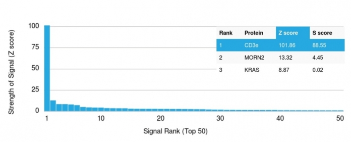
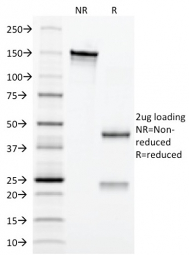
Human Anti-CD3e (T-Cell Marker) Antibody Product Attributes
CD3e (T-Cell Marker) Previously Observed Antibody Staining Patterns
Observed Antibody Staining Data By Tissue Type:
Variations in CD3e antibody staining intensity in immunohistochemistry on tissue sections are present across different anatomical locations. An intense signal was observed in lymphoid tissue in appendix and non-germinal center cells in the lymph node and tonsil. More moderate antibody staining intensity was present in lymphoid tissue in appendix and non-germinal center cells in the lymph node and tonsil. Low, but measureable presence of CD3e could be seen in. We were unable to detect CD3e in other tissues. Disease states, inflammation, and other physiological changes can have a substantial impact on antibody staining patterns. These measurements were all taken in tissues deemed normal or from patients without known disease.
| CD3e (T-Cell Marker) General Information | |
|---|---|
| Alternate Names | |
| CD3 epsilon; CD3 TCR complex; T cell antigen receptor complex epsilon subunit of T3; T-cell surface antigen T3/Leu-4 epsilon chain; T-cell surface glycoprotein CD3 epsilon chain; T3E; TCRE; TiT3 complex | |
| Molecular Weight | |
| 20kDa | |
| Chromosomal Location | |
| Ships on blue ice. | |
| Curated Database and Bioinformatic Data | |
| Gene Symbol | 916 |
| Entrez Gene ID | CD3E |
| UniProt ID(s) | P07766 |
| UniGene ID(s) | Hs3003 |
| COSMIC ID Link(s) | CD3E |
| KEGG Gene ID(s) | hsa:916 |
| General Description of CD3e (T-Cell Marker). | |
| Recognizes the -chain of CD3, which consists of five different polypeptide chains (designated as gamma, delta, epsilon, zeta, and eta) with MW ranging from 16-28kDa. The CD3 complex is closely associated at the lymphocyte cell surface with the T cell antigen receptor (TCR). Reportedly, CD3 complex is involved in signal transduction to the T cell interior following antigen recognition. The CD3 antigen is first detectable in early thymocytes and probably represents one of the earliest signs of commitment to the T cell lineage. In cortical thymocytes, CD3 is predominantly intra-cytoplasmic. However, in medullary thymocytes, it appears on the T cell surface. CD3 antigen is a highly specific marker for T cells, and is present in majority of T cell neoplasms. | |


-150x150.jpg)

There are no reviews yet.