Human Anti-GFAP (Astrocyte & Neural Stem Cell Marker) Antibody Product Attributes
GFAP (Astrocyte & Neural Stem Cell Marker) Previously Observed Antibody Staining Patterns
Observed Subcellular, Organelle Specific Staining Data:
Anti-GFAP antibody staining is expected to be primarily localized to the intermediate filaments. There is variability in either the signal strength or the localization of signal in intermediate filaments from cell to cell.
Observed Antibody Staining Data By Tissue Type:
Variations in GFAP antibody staining intensity in immunohistochemistry on tissue sections are present across different anatomical locations. Low, but measureable presence of GFAP could be seen in. We were unable to detect GFAP in other tissues. Disease states, inflammation, and other physiological changes can have a substantial impact on antibody staining patterns. These measurements were all taken in tissues deemed normal or from patients without known disease.
| GFAP (Astrocyte & Neural Stem Cell Marker) General Information | |
|---|---|
| Alternate Names | |
| Astrocyte or Intermediate Filament Protein, Glial Fibrillary Acidic Protein (GFAP) | |
| Molecular Weight | |
| ~50kDa | |
| Chromosomal Location | |
| Ships on blue ice. | |
| Curated Database and Bioinformatic Data | |
| Gene Symbol | 2670 |
| Entrez Gene ID | GFAP |
| UniProt ID(s) | P14136 |
| UniGene ID(s) | Hs514227 |
| COSMIC ID Link(s) | GFAP |
| KEGG Gene ID(s) | hsa:2670 |
| General Description of GFAP (Astrocyte & Neural Stem Cell Marker). | |
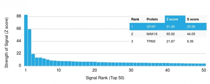
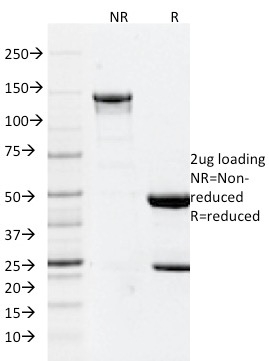
| This MAb recognizes a protein of ~50kDa which is identified as Glial Fibrillary Acidic Protein (GFAP). It shows no cross-reaction with other intermediate filament proteins. GFAP is specifically found in astroglia. GFAP is a very popular marker for localizing benign astrocyte and neoplastic cells of glial origin in the central nervous system. Antibody to GFAP is useful in differentiating primary gliomas from metastatic lesions in the brain and for documenting astrocytic differentiation in tumors outside the CNS. | |


There are no reviews yet.