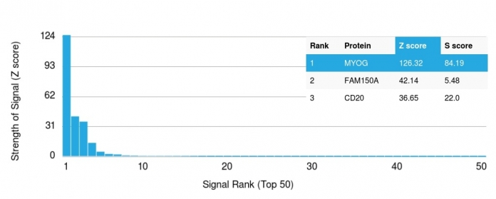
Human Anti-Myogenin (Skeletal Muscle Marker) Antibody Product Attributes
Myogenin (Skeletal Muscle Marker) Previously Observed Antibody Staining Patterns
Observed Subcellular, Organelle Specific Staining Data:
Anti-MYOG antibody staining is expected to be primarily localized to the nucleoplasm. There is variability in either the signal strength or the localization of signal in nucleoplasm from cell to cell.
Observed Antibody Staining Data By Tissue Disease Status:
Tissues from cancer patients, for instance, have their own distinct pattern of Myogenin expression as measured by anti-Myogenin antibody immunohistochemical staining. The average level of expression by tumor is summarized in the table below. The variability row represents patient to patient variability in IHC staining.
| Sample Type | breast cancer | carcinoid | cervical cancer | colorectal cancer | endometrial cancer | glioma | head and neck cancer | liver cancer | lung cancer | lymphoma | melanoma | ovarian cancer | pancreatic cancer | prostate cancer | renal cancer | skin cancer | stomach cancer | testicular cancer | thyroid cancer | urothelial cancer |
|---|---|---|---|---|---|---|---|---|---|---|---|---|---|---|---|---|---|---|---|---|
| Signal Intensity | + | + | – | – | – | + | + | + | – | + | + | ++ | – | + | + | – | – | + | – | – |
| MYOG Variability | ++ | ++ | ++ | + | ++ | ++ | ++ | ++ | + | ++ | ++ | ++ | ++ | ++ | +++ | ++ | ++ | ++ | ++ | ++ |
| Myogenin (Skeletal Muscle Marker) General Information | |
|---|---|
| Alternate Names | |
| bHLHc3, cb553, Class C basic helix-loop-helix protein 3, Myf-4, MYF4, MYOG, Myogenic factor 4 | |
| Molecular Weight | |
| 34kDa | |
| Chromosomal Location | |
| Ships on blue ice. | |
| Curated Database and Bioinformatic Data | |
| Gene Symbol | 4656 |
| Entrez Gene ID | MYOG |
| UniProt ID(s) | P15173 |
| UniGene ID(s) | Hs2830 |
| COSMIC ID Link(s) | MYOG |
| KEGG Gene ID(s) | hsa:4656 |
| General Description of Myogenin (Skeletal Muscle Marker). | |
| Myogenin is a member of the MyoD family of myogenic basic helix-loop-helix (bHLH) transcription factors that also includes MyoD, Myf-5, and MRF4 (also known as herculinor Myf-6). MyoD family members are expressed exclusively in skeletal muscle and play a key role in activating myogenesis by binding to enhancer sequences of muscle-specific genes. The regulatory domain of MyoD is approximately 70 amino acids in length and includes both a basic DNA binding motif and a bHLH dimerization motif. MyoD family members share about 80% amino acid homology in their bHLH motifs. Anti-myogenin labels the nuclei of myoblasts in developing muscle tissue, and is expressed in tumor cell nuclei of rhabdomyosarcoma and some leiomyosarcomas. Positive nuclear staining may occur in Wilms tumor. | |

.jpg)

.jpg)
%20Gel.jpg)


There are no reviews yet.