


![Anti-CD45 Antibody [HI30] - Image 6](https://cdn-enquirebio.pressidium.com/wp-content/uploads/2017/10/enQuire-Bio-QAB46-PE-100Tests-anti-CD45-antibody-10.png)


 PDF Datasheet
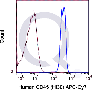
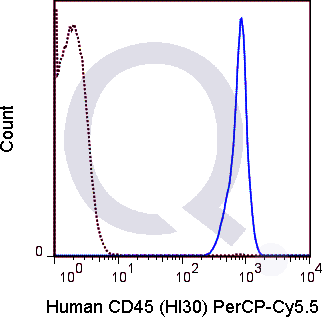
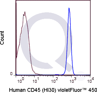
Human Anti-CD45 Antibody Product Attributes
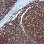
CD45 Previously Observed Antibody Staining Patterns
Observed Antibody Staining Data By Tissue Type:
Variations in CD45 / PTPRC antibody staining intensity in immunohistochemistry on tissue sections are present across different anatomical locations. An intense signal was observed in lymphoid tissue in appendix, hematopoietic cells in the bone marrow, germinal center cells in the lymph node, non-germinal center cells in the lymph node, cells in the red pulp in spleen, cells in the white pulp in spleen, germinal center cells in the tonsil and non-germinal center cells in the tonsil. More moderate antibody staining intensity was present in lymphoid tissue in appendix, hematopoietic cells in the bone marrow, germinal center cells in the lymph node, non-germinal center cells in the lymph node, cells in the red pulp in spleen, cells in the white pulp in spleen, germinal center cells in the tonsil and non-germinal center cells in the tonsil. Low, but measureable presence of CD45 / PTPRC could be seen in. We were unable to detect CD45 / PTPRC in other tissues. Disease states, inflammation, and other physiological changes can have a substantial impact on antibody staining patterns. These measurements were all taken in tissues deemed normal or from patients without known disease.
Observed Antibody Staining Data By Tissue Disease Status:
Tissues from cancer patients, for instance, have their own distinct pattern of CD45 expression as measured by anti-CD45 antibody immunohistochemical staining. The average level of expression by tumor is summarized in the table below. The variability row represents patient to patient variability in IHC staining.
| Sample Type | breast cancer | carcinoid | cervical cancer | colorectal cancer | endometrial cancer | glioma | head and neck cancer | liver cancer | lung cancer | lymphoma | melanoma | ovarian cancer | pancreatic cancer | prostate cancer | renal cancer | skin cancer | stomach cancer | testicular cancer | thyroid cancer | urothelial cancer |
|---|---|---|---|---|---|---|---|---|---|---|---|---|---|---|---|---|---|---|---|---|
| Signal Intensity | – | – | – | – | – | – | – | – | – | +++ | – | – | – | – | – | – | – | – | – | – |
| PTPRC Variability | + | + | + | + | + | + | + | + | + | + | + | + | + | + | + | + | + | + | + | + |
| CD45 General Information | |
|---|---|
| Alternate Names | |
| CD45, PTPRC, Lyt-4, T2, loc, CD45R, Ly-5, B22, Cd45, L-CA, LCA | |
| Curated Database and Bioinformatic Data | |
| Gene Symbol | PTPRC |
| Entrez Gene ID | 5788 |
| Ensemble Gene ID | ENSG00000081237, ENSG00000262418 |
| RefSeq Protein Accession(s) | XP_006711537, XP_006711536, XP_006711535, NP_563578, NP_001254727, NP_002829 |
| RefSeq mRNA Accession(s) | XM_006711473, XM_006711474, NM_080921, NM_080922, NM_001267798, NM_002838, XM_006711472, NR_052021 |
| RefSeq Genomic Accession(s) | NG_007730, NC_000001, NW_003315907, NC_018912 |
| UniProt ID(s) | X6R433, A0A0A0MT22, P08575, M9MML4 |
| UniGene ID(s) | X6R433, A0A0A0MT22, P08575, M9MML4 |
| HGNC ID(s) | 9666 |
| Cosmic ID(s) | PTPRC |
| KEGG Gene ID(s) | hsa:5788 |
| PharmGKB ID(s) | PA34011 |
| General Description of CD45. | |
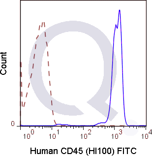
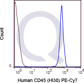
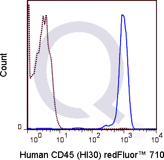
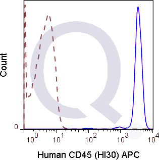
| The HI30 antibody reacts with human CD45, one of the most abundant hematopoietic markers and one that is expressed on all leukocytes (the Leukocyte Common Antigen, LCA). CD45 is a protein tyrosine phosphatase existing in several isoforms, each being generated and expressed in cell-specific patterns. With its broad cell distribution, CD45 is critical for many leukocyte functions, regulating signal transduction and cell activation associated with the T cell receptor, B cell receptor, and IL-2 receptor. Other forms of CD45, with restricted cellular expression, include CD45R (B220), CD45RA, CD45RB, CD45RO and others.The HI30 antibody is widely used as a marker for human CD45 expression on T cells, B cells, monocytes, macrophages, and NK cells. | |
Selected References
Limitations and Warranty
| Size | |
|---|---|
| Tag | APC, APC-Cy7, FITC, PE, PE-Cy7, PerCP-Cy5.5, Qfluor™ 630, Qfluor™ 710, Unconjugated, V450 |
| Buffer and Stabilizer | 10 mM NaH2PO4, 150 mM NaCl, 0.09% NaN3, 0.1% gelatin, pH7.2, 10 mM NaH2PO4, 150 mM NaCl, 0.09% NaN3, pH 7.2 |
| Product Type | |
| Host | |
| Isotype | |
| Applications | |
| Species | |
| Mass Spec Validated? |
Only logged in customers who have purchased this product may leave a review.
Reviews
There are no reviews yet.