
 PDF Datasheet
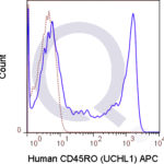
Human, Chimpanzee, and Common Marmoset Anti-CD45RO Antibody Product Attributes
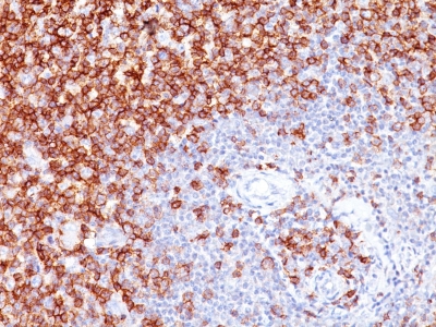
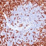
CD45RO Previously Observed Antibody Staining Patterns
Observed Antibody Staining Data By Tissue Type:
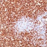
Variations in CD45RO antibody staining intensity in immunohistochemistry on tissue sections are present across different anatomical locations. An intense signal was observed in lymphoid tissue in appendix, hematopoietic cells in the bone marrow, germinal center cells in the lymph node, non-germinal center cells in the lymph node, cells in the red pulp in spleen, cells in the white pulp in spleen, germinal center cells in the tonsil and non-germinal center cells in the tonsil. More moderate antibody staining intensity was present in lymphoid tissue in appendix, hematopoietic cells in the bone marrow, germinal center cells in the lymph node, non-germinal center cells in the lymph node, cells in the red pulp in spleen, cells in the white pulp in spleen, germinal center cells in the tonsil and non-germinal center cells in the tonsil. Low, but measureable presence of CD45RO could be seen in. We were unable to detect CD45RO in other tissues. Disease states, inflammation, and other physiological changes can have a substantial impact on antibody staining patterns. These measurements were all taken in tissues deemed normal or from patients without known disease.
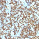
Observed Antibody Staining Data By Tissue Disease Status:
Tissues from cancer patients, for instance, have their own distinct pattern of CD45RO expression as measured by anti-CD45RO antibody immunohistochemical staining. The average level of expression by tumor is summarized in the table below. The variability row represents patient to patient variability in IHC staining.
| Sample Type | breast cancer | carcinoid | cervical cancer | colorectal cancer | endometrial cancer | glioma | head and neck cancer | liver cancer | lung cancer | lymphoma | melanoma | ovarian cancer | pancreatic cancer | prostate cancer | renal cancer | skin cancer | stomach cancer | testicular cancer | thyroid cancer | urothelial cancer |
|---|---|---|---|---|---|---|---|---|---|---|---|---|---|---|---|---|---|---|---|---|
| Signal Intensity | – | – | – | – | – | – | – | – | – | +++ | – | – | – | – | – | – | – | – | – | – |
| PTPRC Variability | + | + | + | + | + | + | + | + | + | + | + | + | + | + | + | + | + | + | + | + |
| CD45RO General Information | |
|---|---|
| Alternate Names | |
| Protein tyrosine phosphatase, receptor type, C, PTPRC | |
| Molecular Weight | |
| 180-185kDa | |
| Chromosomal Location | |
| 1q31.3 | |
| Curated Database and Bioinformatic Data | |
| Gene Symbol | PTPRC |
| Entrez Gene ID | 5788 |
| Ensemble Gene ID | ENSG00000081237, ENSG00000262418 |
| RefSeq Protein Accession(s) | XP_006711537, XP_006711536, XP_006711535, NP_563578, NP_001254727, NP_002829 |
| RefSeq mRNA Accession(s) | XM_006711473, XM_006711474, NM_080921, NM_080922, NM_001267798, NM_002838, XM_006711472, NR_052021 |
| RefSeq Genomic Accession(s) | NG_007730, NC_000001, NW_003315907, NC_018912 |
| UniProt ID(s) | X6R433, A0A0A0MT22, P08575, M9MML4 |
| UniGene ID(s) | X6R433, A0A0A0MT22, P08575, M9MML4 |
| HGNC ID(s) | 9666 |
| Cosmic ID(s) | PTPRC |
| KEGG Gene ID(s) | hsa:5788 |
| PharmGKB ID(s) | PA34011 |
| General Description of CD45RO. | |
| Recognizes a 180-185kDa protein, identified as isoform of leukocyte common antigen (CD45RO) (4th Leucocyte Typing Workshop: Code No. N31). The epitope recognized by this antibody is sensitive to neuraminidase digestion. This antibody reacts with mature activated T-cells, most thymocytes,, a sub-population of resting T-cells within both CD4, CD8 subsets. It shows no reactivity with normal B or natural killer cells, but reacts with granulocytes, monocytes. Reportedly, it is useful to identify T-cell lymphomas, leukemias. It rarely stains NK cells or B-cell lymphomas. | |
Reviews
There are no reviews yet.