
 PDF Datasheet
Human Anti-CD56 / NCAM1 / NKH1 Antibody Product Attributes
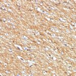
CD56 / NCAM1 / NKH1 Previously Observed Antibody Staining Patterns
Observed Subcellular, Organelle Specific Staining Data:
Anti-NCAM1 antibody staining is expected to be primarily localized to the cytosol and plasma membrane.
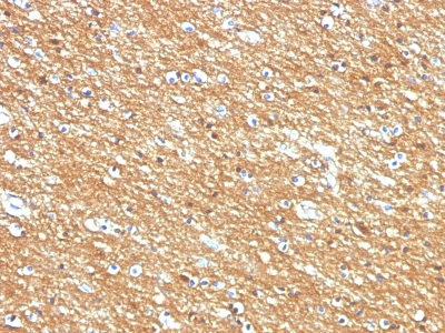
Observed Antibody Staining Data By Tissue Type:
Variations in CD56 / NCAM1 / NKH1 antibody staining intensity in immunohistochemistry on tissue sections are present across different anatomical locations. An intense signal was observed in neuropil in cerebral cortex and peripheral nerve/ganglion in colon. More moderate antibody staining intensity was present in neuropil in cerebral cortex and peripheral nerve/ganglion in colon. Low, but measureable presence of CD56 / NCAM1 / NKH1 could be seen inglial cells in the hippocampus, exocrine glandular cells in the pancreas and smooth muscle cells in the smooth muscle. We were unable to detect CD56 / NCAM1 / NKH1 in other tissues. Disease states, inflammation, and other physiological changes can have a substantial impact on antibody staining patterns. These measurements were all taken in tissues deemed normal or from patients without known disease.
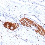
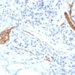
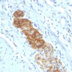
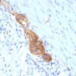
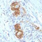
Observed Antibody Staining Data By Tissue Disease Status:
Tissues from cancer patients, for instance, have their own distinct pattern of CD56 / NCAM1 / NKH1 expression as measured by anti-CD56 / NCAM1 / NKH1 antibody immunohistochemical staining. The average level of expression by tumor is summarized in the table below. The variability row represents patient to patient variability in IHC staining.
| Sample Type | breast cancer | carcinoid | cervical cancer | colorectal cancer | endometrial cancer | glioma | head and neck cancer | liver cancer | lung cancer | lymphoma | melanoma | ovarian cancer | pancreatic cancer | prostate cancer | renal cancer | skin cancer | stomach cancer | testicular cancer | thyroid cancer | urothelial cancer |
|---|---|---|---|---|---|---|---|---|---|---|---|---|---|---|---|---|---|---|---|---|
| Signal Intensity | – | – | – | – | – | ++ | – | – | – | – | – | – | – | – | – | – | – | – | – | – |
| NCAM1 Variability | + | ++ | + | + | + | ++ | + | + | + | + | + | ++ | + | + | + | + | + | + | ++ | + |
| CD56 / NCAM1 / NKH1 General Information | |
|---|---|
| Alternate Names | |
| Neural cell adhesion molecule, NCAM, CD56 | |
| Molecular Weight | |
| 180, 145, 125kDa | |
| Chromosomal Location | |
| 11q23.1 | |
| Curated Database and Bioinformatic Data | |
| Gene Symbol | NCAM1 |
| Entrez Gene ID | 4684 |
| Ensemble Gene ID | ENSG00000149294 |
| RefSeq Protein Accession(s) | NP_001229536, NP_851996, NP_001070150, NP_001229537, NP_000606 |
| RefSeq mRNA Accession(s) | NM_001076682, NM_001242607, NM_001242608, NM_181351 NM_000615 |
| RefSeq Genomic Accession(s) | NC_000011, NG_032036, NC_018922 |
| UniProt ID(s) | P13591, A0A087WWD4 |
| UniGene ID(s) | P13591, A0A087WWD4 |
| HGNC ID(s) | 7656 |
| Cosmic ID(s) | NCAM1 |
| KEGG Gene ID(s) | hsa:4684 |
| PharmGKB ID(s) | PA31459 |
| General Description of CD56 / NCAM1 / NKH1. | |
| This MAb reacts with an extracellular domain (close to transmembrane) of CD56/NCAM. Three isoforms of neural cell adhesion molecule (NCAM) are produced by differential splicing of the RNA transcript from a single gene. The 135kDa isoform is the basic molecule, which is glycosylated or sialylated to produce the mature species. Anti-CD56 recognizes two proteins of the neural cell adhesion molecule, the basic molecule expressed on most neuroectodermally derived tissues, neoplasms (e.g. retinoblastoma, medulloblastomas, astrocytomas, neuroblastomas,, small cell carcinomas). It is also expressed on some mesodermally derived tumors (rhabdomyosarcoma). Anti-CD56 plays an important role in the diagnosis of nodal, nasal NK/T-cell lymphomas. | |
Reviews
There are no reviews yet.