
 PDF Datasheet
Human, Mouse (-), Rat (-), Hamster (-), Cow (-), Canine (-), Pig (-), and Sheep (-) Anti-Cytokeratin 18 Antibody Product Attributes
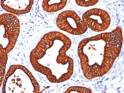
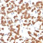
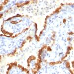
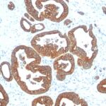
Cytokeratin 18 Previously Observed Antibody Staining Patterns
Observed Subcellular, Organelle Specific Staining Data:
Anti-KRT18 antibody staining is expected to be primarily localized to the cytosol.
Observed Antibody Staining Data By Tissue Type:
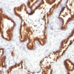
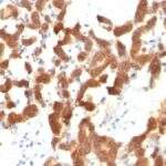
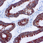
Variations in Cytokeratin 18 antibody staining intensity in immunohistochemistry on tissue sections are present across different anatomical locations. An intense signal was observed in bile duct cells in the liver, cells in the tubules in kidney, exocrine glandular cells in the pancreas, glandular cells in the appendix, breast, cervix, uterine, colon, duodenum, endometrium, epididymis, fallopian tube, gallbladder, parathyroid gland, prostate, rectum, salivary gland, seminal vesicle, small intestine, stomach and thyroid gland, hepatocytes in liver, pneumocytes in lung, respiratory epithelial cells in the bronchus and nasopharynx and trophoblastic cells in the placenta. More moderate antibody staining intensity was present in bile duct cells in the liver, cells in the tubules in kidney, exocrine glandular cells in the pancreas, glandular cells in the appendix, breast, cervix, uterine, colon, duodenum, endometrium, epididymis, fallopian tube, gallbladder, parathyroid gland, prostate, rectum, salivary gland, seminal vesicle, small intestine, stomach and thyroid gland, hepatocytes in liver, pneumocytes in lung, respiratory epithelial cells in the bronchus and nasopharynx and trophoblastic cells in the placenta. Low, but measureable presence of Cytokeratin 18 could be seen in. We were unable to detect Cytokeratin 18 in other tissues. Disease states, inflammation, and other physiological changes can have a substantial impact on antibody staining patterns. These measurements were all taken in tissues deemed normal or from patients without known disease.
Observed Antibody Staining Data By Tissue Disease Status:
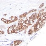
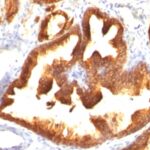
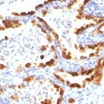
Tissues from cancer patients, for instance, have their own distinct pattern of Cytokeratin 18 expression as measured by anti-Cytokeratin 18 antibody immunohistochemical staining. The average level of expression by tumor is summarized in the table below. The variability row represents patient to patient variability in IHC staining.
| Sample Type | breast cancer | carcinoid | cervical cancer | colorectal cancer | endometrial cancer | glioma | head and neck cancer | liver cancer | lung cancer | lymphoma | melanoma | ovarian cancer | pancreatic cancer | prostate cancer | renal cancer | skin cancer | stomach cancer | testicular cancer | thyroid cancer | urothelial cancer |
|---|---|---|---|---|---|---|---|---|---|---|---|---|---|---|---|---|---|---|---|---|
| Signal Intensity | ++ | +++ | ++ | +++ | +++ | – | – | +++ | +++ | – | – | +++ | +++ | +++ | ++ | – | +++ | – | +++ | ++ |
| KRT18 Variability | ++ | + | +++ | ++ | + | + | ++ | ++ | ++ | + | + | + | + | + | ++ | + | + | ++ | + | ++ |
| Cytokeratin 18 General Information | |
|---|---|
| Alternate Names | |
| Keratin 18 | |
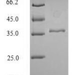
| Molecular Weight | |
| 45kDa | |
| Chromosomal Location | |
| 12q13.13 | |
| Curated Database and Bioinformatic Data | |
| Gene Symbol | KRT18 |
| Entrez Gene ID | 3875 |
| Ensemble Gene ID | ENSG00000111057 |
| RefSeq Protein Accession(s) | NP_954657, NP_000215 |
| RefSeq mRNA Accession(s) | NM_199187, NM_000224 |
| RefSeq Genomic Accession(s) | NC_000012, NG_008351, NG_008402, NC_018923 |
| UniProt ID(s) | P05783, A0A024RAY2 |
| UniGene ID(s) | P05783, A0A024RAY2 |
| HGNC ID(s) | 6430 |
| Cosmic ID(s) | KRT18 |
| KEGG Gene ID(s) | hsa:3875 |
| PharmGKB ID(s) | PA30217 |
| General Description of Cytokeratin 18. | |
| This MAb reacts with a wide variety of simple epithelia. It does not react with stratified squamous epithelia. It reacts with epithelial tumors of the gastrointestinal tract, lung, breast, pancreas, ovary,, thyroid. Cytokeratin 18, which belongs to the type A (acidic) subfamily of low molecular weight keratins, exists in combination with cytokeratin 8. It is reported that tissues from gastrointestinal tract are positive for both cytokeratin 8, 18 but do not contain cytokeratin 14. Tissues from gastrointestinal tract, respiratory tract, urogenital tract, as well as endocrine, exocrine tissues, mesothelial cells are positive for cytokeratin 18. | |
![Analysis of Mass Spec data (dashed-line) of fractions stained with Cytokeratin 18 <a href="https://enquirebio.com/validation-project-details/" target="_blank">MS-QAVA™ monoclonal antibody</a> [Clone: B23.1] (solid-line), reveals that less than 13.5% of signal is attributable to non-specific binding of anti-Cytokeratin 18 [Clone B23.1] to targets other than KRT18 protein. Even frequently cited antibodies have much greater non-specific interactions, averaging over 30%. Data in image is from analysis in A431, RT4 and MCF7 cells.](https://cdn-enquirebio.pressidium.com/wp-content/uploads/2017/10/enQuire-Bio-3875-MSM9-P1-anti-Cytokeratin-18-antibody-150x150.png)

Reviews
There are no reviews yet.