
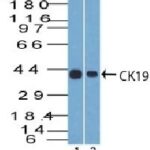
![Analysis of Mass Spec data (dashed-line) of fractions stained with Cytokeratin 19 MS-QAVA™ monoclonal antibody [Clone: KRT19/799] (solid-line), reveals that less than 2.3% of signal is attributable to non-specific binding of anti-Cytokeratin 19 [Clone KRT19/799] to targets other than KRT19 protein. Even frequently cited antibodies have much greater non-specific interactions, averaging over 30%. Data in image is from analysis in Jurkat, U202 and HeLa cells.](https://cdn-enquirebio.pressidium.com/wp-content/uploads/2017/10/enQuire-Bio-3880-MSM4-P1-anti-Cytokeratin-19-antibody.png)
 PDF Datasheet
Human Anti-Cytokeratin 19 Antibody Product Attributes
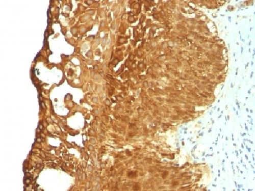
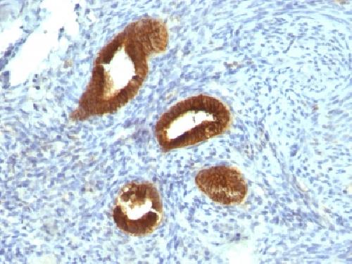
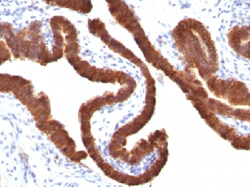
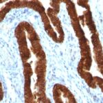
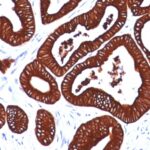
Cytokeratin 19 Previously Observed Antibody Staining Patterns
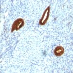
Observed Subcellular, Organelle Specific Staining Data:
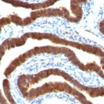
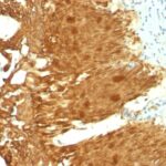
Variations in Cytokeratin 19 antibody staining intensity in immunohistochemistry on tissue sections are present across different anatomical locations. An intense signal was observed in bile duct cells in the liver, cells in the tubules in kidney, exocrine glandular cells in the pancreas, glandular cells in the appendix, breast, cervix, uterine, colon, duodenum, endometrium, epididymis, fallopian tube, gallbladder, prostate, rectum, salivary gland, seminal vesicle, small intestine, stomach and thyroid gland, pneumocytes in lung, respiratory epithelial cells in the bronchus and nasopharynx, squamous epithelial cells in the tonsil, trophoblastic cells in the placenta and urothelial cells in the urinary bladder. More moderate antibody staining intensity was present in bile duct cells in the liver, cells in the tubules in kidney, exocrine glandular cells in the pancreas, glandular cells in the appendix, breast, cervix, uterine, colon, duodenum, endometrium, epididymis, fallopian tube, gallbladder, prostate, rectum, salivary gland, seminal vesicle, small intestine, stomach and thyroid gland, pneumocytes in lung, respiratory epithelial cells in the bronchus and nasopharynx, squamous epithelial cells in the tonsil, trophoblastic cells in the placenta and urothelial cells in the urinary bladder. Low, but measureable presence of Cytokeratin 19 could be seen inepidermal cells in the skin, squamous epithelial cells in the cervix and uterine and oral mucosa. We were unable to detect Cytokeratin 19 in other tissues. Disease states, inflammation, and other physiological changes can have a substantial impact on antibody staining patterns. These measurements were all taken in tissues deemed normal or from patients without known disease.
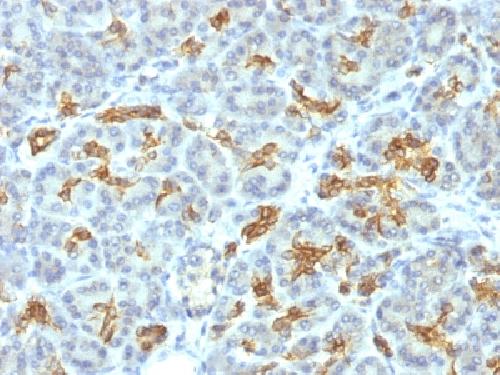
Observed Antibody Staining Data By Tissue Type:
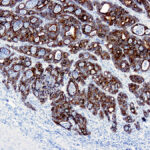
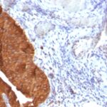
Tissues from cancer patients, for instance, have their own distinct pattern of Cytokeratin 19 expression as measured by anti-Cytokeratin 19 antibody immunohistochemical staining. The average level of expression by tumor is summarized in the table below. The variability row represents patient to patient variability in IHC staining.
| Sample Type | breast cancer | carcinoid | cervical cancer | colorectal cancer | endometrial cancer | glioma | head and neck cancer | liver cancer | lung cancer | lymphoma | melanoma | ovarian cancer | pancreatic cancer | prostate cancer | renal cancer | skin cancer | stomach cancer | testicular cancer | thyroid cancer | urothelial cancer |
|---|---|---|---|---|---|---|---|---|---|---|---|---|---|---|---|---|---|---|---|---|
| Signal Intensity | +++ | +++ | +++ | +++ | +++ | – | + | – | +++ | – | – | +++ | +++ | +++ | + | ++ | +++ | – | +++ | +++ |
| KRT19 Variability | + | + | + | + | + | + | ++ | ++ | + | + | + | + | + | ++ | +++ | ++ | + | + | ++ | + |
| Cytokeratin 19 General Information | |
|---|---|
| Alternate Names | |
| Keratin, type I cytoskeletal 19, cytokeratin-19, CK-19, keratin-19, K19, KRT19 | |
| Molecular Weight | |
| 40kDa | |
| Chromosomal Location | |
| 17q21.2 | |
| Curated Database and Bioinformatic Data | |
| Gene Symbol | KRT19 |
| Entrez Gene ID | 3880 |
| Ensemble Gene ID | ENSG00000171345 |
| RefSeq Protein Accession(s) | NP_002267 |
| RefSeq mRNA Accession(s) | NM_002276 |
| RefSeq Genomic Accession(s) | NC_000017, NC_018928, NG_012285 |
| UniProt ID(s) | P08727 |
| UniGene ID(s) | P08727 |
| HGNC ID(s) | 6436 |
| Cosmic ID(s) | KRT19 |
| KEGG Gene ID(s) | hsa:3880 |
| PharmGKB ID(s) | PA30225 |
| General Description of Cytokeratin 19. | |
| Recognizes a protein of 40kDa, identified as cytokeratin-19 (CK19), which is expressed in sweat gland, mammary gland ductal, secretory cells, bile ducts, gastrointestinal tract, bladder urothelium, oral epithelia, esophagus,, ectocervical epithelium. Anti-CK19 reacts with a wide variety of epithelial malignancies including adenocarcinomas of the colon, stomach, pancreas, biliary tract, liver,, breast. Perhaps the most useful application is the identification of thyroid carcinoma of the papillary type, although 50%-60% of follicular carcinomas are also labeled. Anti-CK19 is a useful marker for detection of tumor cells in lymph nodes, peripheral blood, bone marrow, breast cancer. | |

Reviews
There are no reviews yet.