
 PDF Datasheet
Human Anti-Estrogen Receptor Antibody Product Attributes
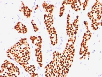
Estrogen Receptor Previously Observed Antibody Staining Patterns
Observed Subcellular, Organelle Specific Staining Data:
Anti-ESR1 antibody staining is expected to be primarily localized to the nucleus and vesicles.
Observed Antibody Staining Data By Tissue Type:
Variations in Estrogen Receptor antibody staining intensity in immunohistochemistry on tissue sections are present across different anatomical locations. An intense signal was observed in cells in the endometrial stroma in endometrium, glandular cells in the cervix, uterine, endometrium and fallopian tube, squamous epithelial cells in the cervix and uterine. More moderate antibody staining intensity was present in cells in the endometrial stroma in endometrium, glandular cells in the cervix, uterine, endometrium and fallopian tube, squamous epithelial cells in the cervix and uterine. Low, but measureable presence of Estrogen Receptor could be seen insmooth muscle cells in the smooth muscle. We were unable to detect Estrogen Receptor in other tissues. Disease states, inflammation, and other physiological changes can have a substantial impact on antibody staining patterns. These measurements were all taken in tissues deemed normal or from patients without known disease.
Observed Antibody Staining Data By Tissue Disease Status:
Tissues from cancer patients, for instance, have their own distinct pattern of Estrogen Receptor expression as measured by anti-Estrogen Receptor antibody immunohistochemical staining. The average level of expression by tumor is summarized in the table below. The variability row represents patient to patient variability in IHC staining.
| Sample Type | breast cancer | carcinoid | cervical cancer | colorectal cancer | endometrial cancer | glioma | head and neck cancer | liver cancer | lung cancer | lymphoma | melanoma | ovarian cancer | pancreatic cancer | prostate cancer | renal cancer | skin cancer | stomach cancer | testicular cancer | thyroid cancer | urothelial cancer |
|---|---|---|---|---|---|---|---|---|---|---|---|---|---|---|---|---|---|---|---|---|
| Signal Intensity | +++ | – | – | – | + | – | – | – | – | – | – | – | – | – | – | – | – | – | – | – |
| ESR1 Variability | ++ | ++ | + | + | ++ | + | + | + | + | + | + | ++ | + | + | + | + | + | + | + | + |
| Estrogen Receptor General Information | |
|---|---|
| Alternate Names | |
| Estrogen receptor alpha, ER-?, ERalpha, ER alpha, ESR1 | |
| Molecular Weight | |
| ~67kDa | |
| Chromosomal Location | |
| 6q25.1 | |
| Curated Database and Bioinformatic Data | |
| Gene Symbol | ESR1 |
| Entrez Gene ID | 2099 |
| Ensemble Gene ID | ENSG00000091831 |
| RefSeq Protein Accession(s) | XP_011533846, XP_016865866, XP_016865871, NP_001315029, XP_011533845, XP_016865867, XP_016865870, XP_016865872, XP_006715438, XP_016865865, XP_016865869, NP_000116, NP_001116212, XP_011533849, NP_001116213, XP_011533847, XP_011533851, XP_016865868, XP_006715437, NP_001116214, NP_001278159, NP_001278170 |
| RefSeq mRNA Accession(s) | XM_011535547, NM_000125, XM_011535544, XM_017010376 NM_001122742, XM_017010380, XM_017010381, XM_011535543, XM_017010378, XR_001743222, NM_001328100, XM_011535549, XM_017010377, XM_017010379, XM_017010382, NM_001122740, NM_001122741, NM_001291241, XM_011535545, XM_017010383, XM_006715374, XM_006715375, XR_001743223, NM_001291230 |
| RefSeq Genomic Accession(s) | NC_000006, NC_018917, NG_008493 |
| UniProt ID(s) | A8KAF4, A0A125SXW3, Q9UBT1, H0Y4W6, P03372, G4XH65 |
| UniGene ID(s) | A8KAF4, A0A125SXW3, Q9UBT1, H0Y4W6, P03372, G4XH65 |
| HGNC ID(s) | 3467 |
| Cosmic ID(s) | ESR1 |
| KEGG Gene ID(s) | hsa:2099 |
| PharmGKB ID(s) | PA156 |
| General Description of Estrogen Receptor. | |
| This MAb is specific to ER alpha, shows minimal cross-reaction with other members of the family. ER is an important regulator of growth, differentiation in the mammary gland. Presence of ER in breast tumors indicates an increased likelihood of response to anti-estrogen (e.g. tamoxifen) therapy. This MAb is excellent for staining of formalin-fixed, paraffin-embedded breast carcinomas. | |
Reviews
There are no reviews yet.