
 PDF Datasheet
Human, Monkey, Cow, Pig, Mouse, and Fish Anti-Heparan Sulfate Proteoglycan / Perlecan Antibody Product Attributes
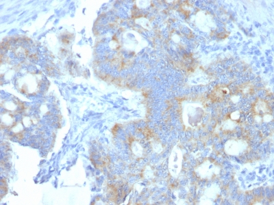
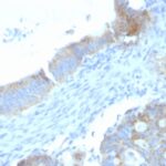
Heparan Sulfate Proteoglycan / Perlecan Previously Observed Antibody Staining Patterns
Observed Subcellular, Organelle Specific Staining Data:
Anti-HSPG2 antibody staining is expected to be primarily localized to the cytosol, nucleoplasm and plasma membrane.
Observed Antibody Staining Data By Tissue Type:
Variations in Heparan Sulfate Proteoglycan / Perlecan antibody staining intensity in immunohistochemistry on tissue sections are present across different anatomical locations. Low, but measureable presence of Heparan Sulfate Proteoglycan / Perlecan could be seen inadipocytes in mesenchymal tissue, cells in the tubules in kidney, decidual cells in the placenta, endothelial cells in the cerebral cortex, epidermal cells in the skin, exocrine glandular cells in the pancreas, fibroblasts in skin, germinal center cells in the tonsil, glandular cells in the appendix, breast, cervix, uterine, colon, gallbladder, parathyroid gland, rectum and stomach, hematopoietic cells in the bone marrow, hepatocytes in liver, islets of Langerhans in pancreas, Leydig cells in the testis, lymphoid tissue in appendix, macrophages in lung, melanocytes in skin, myoepithelial cells in the breast, non-germinal center cells in the lymph node and tonsil, ovarian stroma cells in the ovary, respiratory epithelial cells in the bronchus, smooth muscle cells in the smooth muscle, squamous epithelial cells in the oral mucosa and tonsil and urothelial cells in the urinary bladder. We were unable to detect Heparan Sulfate Proteoglycan / Perlecan in other tissues. Disease states, inflammation, and other physiological changes can have a substantial impact on antibody staining patterns. These measurements were all taken in tissues deemed normal or from patients without known disease.
Observed Antibody Staining Data By Tissue Disease Status:
Tissues from cancer patients, for instance, have their own distinct pattern of Heparan Sulfate Proteoglycan / Perlecan expression as measured by anti-Heparan Sulfate Proteoglycan / Perlecan antibody immunohistochemical staining. The average level of expression by tumor is summarized in the table below. The variability row represents patient to patient variability in IHC staining.
| Sample Type | breast cancer | carcinoid | cervical cancer | colorectal cancer | endometrial cancer | glioma | head and neck cancer | liver cancer | lung cancer | lymphoma | melanoma | ovarian cancer | pancreatic cancer | prostate cancer | renal cancer | skin cancer | stomach cancer | testicular cancer | thyroid cancer | urothelial cancer |
|---|---|---|---|---|---|---|---|---|---|---|---|---|---|---|---|---|---|---|---|---|
| Signal Intensity | ++ | + | ++ | ++ | ++ | + | ++ | + | ++ | ++ | ++ | + | ++ | + | ++ | ++ | + | ++ | ++ | ++ |
| HSPG2 Variability | ++ | ++ | ++ | + | ++ | ++ | ++ | ++ | ++ | ++ | ++ | ++ | ++ | ++ | ++ | ++ | ++ | ++ | ++ | + |
| Heparan Sulfate Proteoglycan / Perlecan General Information | |
|---|---|
| Alternate Names | |
| Perlecan, Basement membrane-specific heparan sulfate proteoglycan core protein, HSPG, heparan sulfate proteoglycan 2, HSPG2 | |
| Molecular Weight | |
| >400kDa | |
| Chromosomal Location | |
| 1p36.1-p34 | |
| Curated Database and Bioinformatic Data | |
| Gene Symbol | HSPG2 |
| Entrez Gene ID | 3339 |
| Ensemble Gene ID | ENSG00000142798 |
| RefSeq Protein Accession(s) | XP_016856611, NP_001278789, XP_011539620, XP_016856609, NP_005520, XP_016856610 |
| RefSeq mRNA Accession(s) | XM_017001120, NM_005529, NM_001291860, XM_017001121, XM_017001122, XM_011541318, |
| RefSeq Genomic Accession(s) | NC_018912, NC_000001, NG_016740 |
| UniProt ID(s) | P98160 |
| UniGene ID(s) | P98160 |
| HGNC ID(s) | 5273 |
| Cosmic ID(s) | HSPG2 |
| KEGG Gene ID(s) | hsa:3339 |
| PharmGKB ID(s) | PA29537 |
| General Description of Heparan Sulfate Proteoglycan / Perlecan. | |
| This MAb specifically precipitates heterogeneous material of high MW, identified as perlecan, a major heparan-sulfate proteoglycan (HSPG) within all basement membranes, cell surfaces. It does not cross-react with laminin, fibronectin, or dermatran sulfate proteoglycan. Because of perlecan s strategic location, ability to store, protect growth factors, it has been strongly implicated in the control of tumor cell growth, metastatic behavior. Perlecan possesses angiogenic, growth-promoting attributes primarily by acting as a co-receptor for basic fibroblast growth factor (FGF-2). Suppression of perlecan causes substantial inhibition of neoplastic growth, neovascularization. Thus, perlecan is a potent inducer of neoplasm growth, angiogenesis in vivo, therapeutic interventions targeting this key modulator of tumor progression may improve neoplastic treatment. | |
Reviews
There are no reviews yet.