
 PDF Datasheet
Human, Mouse, Rat, and Cow Anti-p40 Antibody Product Attributes
p40 Previously Observed Antibody Staining Patterns
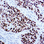
Observed Subcellular, Organelle Specific Staining Data:
Anti-TP63 antibody staining is expected to be primarily localized to the nucleoplasm.
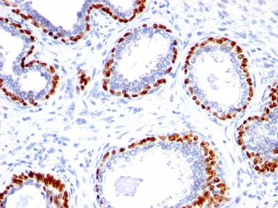
Observed Antibody Staining Data By Tissue Type:
Variations in p40 antibody staining intensity in immunohistochemistry on tissue sections are present across different anatomical locations. An intense signal was observed in myoepithelial cells in the breast, respiratory epithelial cells in the bronchus, squamous epithelial cells in the cervix, uterine, glandular cells in the epididymis, squamous epithelial cells in the esophagus, respiratory epithelial cells in the nasopharynx, squamous epithelial cells in the oral mucosa, keratinocytes in skin, Langerhans in skin, melanocytes in skin, epidermal cells in the skin, squamous epithelial cells in the tonsil, urothelial cells in the urinary bladder and squamous epithelial cells in the vagina. More moderate antibody staining intensity was present in myoepithelial cells in the breast, respiratory epithelial cells in the bronchus, squamous epithelial cells in the cervix, uterine, glandular cells in the epididymis, squamous epithelial cells in the esophagus, respiratory epithelial cells in the nasopharynx, squamous epithelial cells in the oral mucosa, keratinocytes in skin, Langerhans in skin, melanocytes in skin, epidermal cells in the skin, squamous epithelial cells in the tonsil, urothelial cells in the urinary bladder and squamous epithelial cells in the vagina. Low, but measureable presence of p40 could be seen inglandular cells in the breast, cervix, uterine. We were unable to detect p40 in other tissues. Disease states, inflammation, and other physiological changes can have a substantial impact on antibody staining patterns. These measurements were all taken in tissues deemed normal or from patients without known disease.
Observed Antibody Staining Data By Tissue Disease Status:
Tissues from cancer patients, for instance, have their own distinct pattern of p40 expression as measured by anti-p40 antibody immunohistochemical staining. The average level of expression by tumor is summarized in the table below. The variability row represents patient to patient variability in IHC staining.
| Sample Type | breast cancer | carcinoid | cervical cancer | colorectal cancer | endometrial cancer | glioma | head and neck cancer | liver cancer | lung cancer | lymphoma | melanoma | ovarian cancer | pancreatic cancer | prostate cancer | renal cancer | skin cancer | stomach cancer | testicular cancer | thyroid cancer | urothelial cancer |
|---|---|---|---|---|---|---|---|---|---|---|---|---|---|---|---|---|---|---|---|---|
| Signal Intensity | + | – | ++ | – | – | – | +++ | – | ++ | – | – | + | – | – | – | +++ | – | + | – | +++ |
| TP63 Variability | + | ++ | ++ | ++ | ++ | + | + | ++ | ++ | + | ++ | ++ | + | + | + | + | + | ++ | ++ | + |
| p40 General Information | |
|---|---|
| Alternate Names | |
| Tumor protein p63 transformation-related protein 63, TP63, p63 | |
| Molecular Weight | |
| 40kDa | |
| Chromosomal Location | |
| 3q28 | |
| Curated Database and Bioinformatic Data | |
| Gene Symbol | TP63 |
| Entrez Gene ID | 8626 |
| Ensemble Gene ID | ENSG00000073282 |
| RefSeq Protein Accession(s) | NP_003713, XP_016862876, NP_001108451, NP_001108454, XP_005247901, NP_001108450, NP_001316073, NP_001316074, NP_001316077, NP_001108452, XP_005247900, NP_001316075, NP_001316079, XP_011511553, XP_011511554, NP_001108453, NP_001316078, NP_001316893 |
| RefSeq mRNA Accession(s) | XM_005247844, NM_001114978, NM_001114980, NM_001329145, NM_001329146, XM_017007387, NM_001114979, XM_005247843, NM_001114982, NM_001329144, NM_001329964, XM_011513252, NM_001329149, XM_011513251, NM_003722, NM_001329148 NM_001114981, NM_001329150 |
| RefSeq Genomic Accession(s) | NC_000003, NG_007550, NC_018914, |
| UniProt ID(s) | B7Z8X6, A0A0S2Z4N5, A0A141PNN4, A0A141PNN3, Q9H3D4, C9D7D0, A0A0S2Z4N6 |
| UniGene ID(s) | B7Z8X6, A0A0S2Z4N5, A0A141PNN4, A0A141PNN3, Q9H3D4, C9D7D0, A0A0S2Z4N6 |
| HGNC ID(s) | 15979 |
| Cosmic ID(s) | TP63 |
| KEGG Gene ID(s) | hsa:8626 |
| PharmGKB ID(s) | PA162406776 |
| General Description of p40. | |
| p40 (p63 delta) is a marker recently determined to be highly specific for squamous basal cells in the immunohistochemistry (IHC) application. The current more routinely recommended marker, p63, appears to have less specificity compared to p40, especially on squamous cell tumors. The ability to differentiate between lung adenocarcinoma vs. squamous cell carcinoma is difficult, has bearing on the different therapeutic avenues for each subtype treatment. p63 antibodys ability to distinguish between the tumor types appears to be inferior when compared to p40. The ability to utilize an antibody probe for p40 as a squamous cell marker bolsters its use for future sub-classification of lung cancers, especially by immunohistochemical techniques. | |
Reviews
There are no reviews yet.