
 PDF Datasheet
Human Anti-PD1 / PDCD1 / CD279 Antibody Product Attributes
Species: Human
Tested Applications: Flow Cytometry, Immunofluorescence, Immunohistochemistry (IHC).
Application Notes: Flow Cytometry (0.5-1ug of antibody/million cells in 0.1ml), Immunofluorescence (1-2ug of antibody/ml), Immunohistochemistry (IHC) (Formalin-fixed) (0.5-1ug of antibody/ml for 30 minutes at RT)
Clonality: Monoclonal
Anti-PD1 / PDCD1 / CD279 Antibody Clone: PDCD1/922
Clone PDCD1/922 Host and Isotype: Mouse IgG1 kappa
Anti-Human PD1 / PDCD1 / CD279 Positive Control Sample: TY cells or Tonsil.
Cellular Localization of Antibody Cell Surface & Cytoplasmic
Buffer and Stabilizer: 10mM PBS with 0.05% BSA & 0.05% azide.
Antibody Concentration: 200ug/ml
Antibody Purification Method:Protein A/G Purified
Immunogen: Recombinant full-length human PDCD1 protein
Storage Conditions: Store at 2 to 8° C (refrigerate). Stable for 24 months when properly stored.
PD1 / PDCD1 / CD279 Previously Observed Antibody Staining Patterns
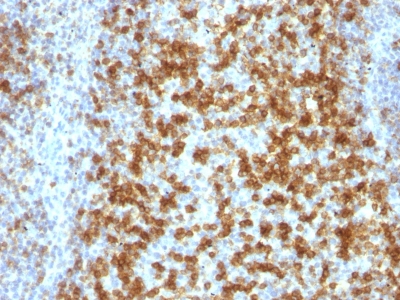
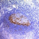
Observed Antibody Staining Data By Tissue Type:
Variations in PD1 / PDCD1 / CD279 antibody staining intensity in immunohistochemistry on tissue sections are present across different anatomical locations. An intense signal was observed in germinal center cells in the tonsil. More moderate antibody staining intensity was present in germinal center cells in the tonsil. Low, but measureable presence of PD1 / PDCD1 / CD279 could be seen in cells in the red pulp in spleen. We were unable to detect PD1 / PDCD1 / CD279 in other tissues. Disease states, inflammation, and other physiological changes can have a substantial impact on antibody staining patterns. These measurements were all taken in tissues deemed normal or from patients without known disease.
| PD1 / PDCD1 / CD279 General Information | |
|---|---|
| Alternate Names | |
| Programmed cell death protein 1, PD-1, PDCD-1, PD1, CD279, cluster of differentiation 279 | |
| Molecular Weight | |
| 55kDa | |
| Chromosomal Location | |
| 2q37.3 | |
| Curated Database and Bioinformatic Data | |
| Gene Symbol | PDCD1 |
| Entrez Gene ID | 5133 |
| Ensemble Gene ID | ENSG00000276977, ENSG00000188389 |
| RefSeq Protein Accession(s) | NP_005009, XP_006712636, XP_016859782 |
| RefSeq mRNA Accession(s) | NM_005018, XM_006712573, XM_017004293 |
| RefSeq Genomic Accession(s) | NG_187527, NC_018913, NC_000002, NG_012110 |
| UniProt ID(s) | Q15116, A0A0M3M0G7 |
| UniGene ID(s) | Q15116, A0A0M3M0G7 |
| HGNC ID(s) | 8760 |
| Cosmic ID(s) | PDCD1 |
| KEGG Gene ID(s) | hsa:5133 |
| PharmGKB ID(s) | PA33110 |
| General Description of PD1 / PDCD1 / CD279. | |
| PDCD-1 (programmed cell death-1 protein), also designated CD279, is a type I transmembrane receptor, a member of the immunoglobin gene superfamily. It is expressed on activated T-cells, B-cells,, myeloid cells. Anti-PDCD-1 is a marker of angioimmunoblastic lymphoma, suggests a unique cell of origin for this neoplasm. Unlike CD10, BCL6, PDCD-1 is expressed by few B-cells, so anti-PDCD-1 may be a more specific, useful diagnostic marker in angioimmunoblastic lymphoma. In addition, PDCD-1 expression provides evidence that angioimmunoblastic lymphoma is a neoplasm derived from germinal center-associated T-cells. | |
Limitations and Warranty
enQuire Bio’s PD1 / PDCD1 / CD279 Anti-Human Monoclonal is available for Research Use Only. This antibody is guaranteed to work for a period of two years when properly stored.

Reviews
There are no reviews yet.