
 PDF Datasheet
Human, Monkey, Canine, Pig, Cow (-), Cat (-), Mouse (-), and Rat (-) Anti-AFP Antibody Product Attributes
AFP Previously Observed Antibody Staining Patterns
Observed Subcellular, Organelle Specific Staining Data:
Anti-AFP antibody staining is expected to be primarily localized to the cytosol.
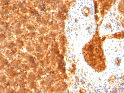
Observed Antibody Staining Data By Tissue Type:
Variations in AFP antibody staining intensity in immunohistochemistry on tissue sections are present across different anatomical locations. Low, but measureable presence of AFP could be seen inglandular cells in the prostate, hepatocytes in liver and trophoblastic cells in the placenta. We were unable to detect AFP in other tissues. Disease states, inflammation, and other physiological changes can have a substantial impact on antibody staining patterns. These measurements were all taken in tissues deemed normal or from patients without known disease.
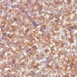
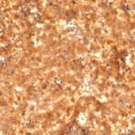
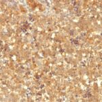
Observed Antibody Staining Data By Tissue Disease Status:
Tissues from cancer patients, for instance, have their own distinct pattern of AFP expression as measured by anti-AFP antibody immunohistochemical staining. The average level of expression by tumor is summarized in the table below. The variability row represents patient to patient variability in IHC staining.
| Sample Type | breast cancer | carcinoid | cervical cancer | colorectal cancer | endometrial cancer | glioma | head and neck cancer | liver cancer | lung cancer | lymphoma | melanoma | ovarian cancer | pancreatic cancer | prostate cancer | renal cancer | skin cancer | stomach cancer | testicular cancer | thyroid cancer | urothelial cancer |
|---|---|---|---|---|---|---|---|---|---|---|---|---|---|---|---|---|---|---|---|---|
| Signal Intensity | + | + | + | + | + | + | + | ++ | + | + | + | ++ | + | + | + | + | + | ++ | + | + |
| AFP Variability | + | ++ | + | + | ++ | ++ | ++ | ++ | ++ | + | ++ | ++ | ++ | ++ | ++ | + | + | ++ | + | ++ |
| AFP General Information | |
|---|---|
| Alternate Names | |
| Alpha-fetoprotein, AFP, ?-fetoprotein | |
| Molecular Weight | |
| 70kDa | |
| Chromosomal Location | |
| 4q13.3 | |
| Curated Database and Bioinformatic Data | |
| Gene Symbol | AFP |
| Entrez Gene ID | 174 |
| Ensemble Gene ID | ENSG00000081051 |
| RefSeq Protein Accession(s) | NP_001125, NP_001341646 |
| RefSeq mRNA Accession(s) | NM_001134, NM_001354717 |
| RefSeq Genomic Accession(s) | NC_000004, NG_023028, NC_018915 |
| UniProt ID(s) | P02771 |
| UniGene ID(s) | P02771 |
| HGNC ID(s) | 317 |
| Cosmic ID(s) | AFP |
| KEGG Gene ID(s) | hsa:174 |
| PharmGKB ID(s) | PA24614 |
| General Description of AFP. | |
| It recognizes an oncofetal glycoprotein with a single chain of 70kDa, which is identified as alpha fetoprotein (AFP). This MAb is highly specific to AFP, shows no cross-reaction with other oncofetal antigens or serum albumin. AFP is normally synthesized in the liver, intestinal tract,, yolk sac of the fetus. Antibody to AFP has been shown to be useful in detecting hepatocellular carcinomas (HCC), germ cell neoplasms, especially yolk sac tumors. | |

Reviews
There are no reviews yet.