
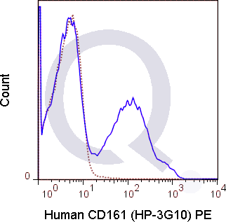
![Human PBMCs were stained with 5 uL Anti-human CD3 antibody [clone OKT3] conjugated to APC (solid line) or 0.25 ug APC Mouse IgG1 isotype control (dashed line). Flow Cytometry Data from 10,000 events.](https://cdn-enquirebio.pressidium.com/wp-content/uploads/2017/10/enQuire-Bio-QAB60-APC-100Tests-anti-CD161-antibody-10.png)
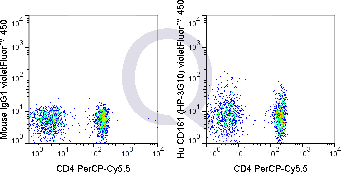
![Anti-CD161 Antibody [HP-3G10] - Image 3](https://cdn-enquirebio.pressidium.com/wp-content/uploads/2017/10/enQuire-Bio-QAB60-F-100Tests-anti-CD161-antibody-10.png)

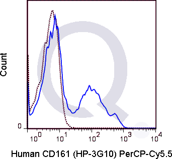
![Human PBMCs were stained with 5 uL Anti-human CD3 antibody [clone OKT3] conjugated to APC (solid line) or 0.25 ug APC Mouse IgG1 isotype control (dashed line). Flow Cytometry Data from 10,000 events.](https://cdn-enquirebio.pressidium.com/wp-content/uploads/2017/11/enQuire-Bio-QAB60-APC-100Tests-anti-CD161-antibody-9.png)


 PDF Datasheet
Human Anti-CD161 Antibody Product Attributes
CD161 Previously Observed Antibody Staining Patterns
Observed Antibody Staining Data By Tissue Type:
Variations in CD161 antibody staining intensity in immunohistochemistry on tissue sections are present across different anatomical locations. An intense signal was observed in cells in the red pulp in spleen, cells in the tubules in kidney, fibroblasts in skin, glandular cells in the epididymis and prostate, hematopoietic cells in the bone marrow and myocytes in heart muscle. More moderate antibody staining intensity was present in cells in the red pulp in spleen, cells in the tubules in kidney, fibroblasts in skin, glandular cells in the epididymis and prostate, hematopoietic cells in the bone marrow and myocytes in heart muscle. Low, but measureable presence of CD161 could be seen in cells in the granular layer in cerebellum, cells in the molecular layer in cerebellum, cells in the white pulp in spleen, endothelial cells in the colon, fibroblasts in mesenchymal tissue, germinal center cells in the tonsil, glandular cells in the cervix, uterine, endometrium, salivary gland and thyroid gland, glial cells in the hippocampus, lymphoid tissue in appendix, neuronal cells in the hippocampus, neuropil in cerebral cortex, peripheral nerve in mesenchymal tissue, peripheral nerve/ganglion in colon, pneumocytes in lung and respiratory epithelial cells in the nasopharynx. We were unable to detect CD161 in other tissues. Disease states, inflammation, and other physiological changes can have a substantial impact on antibody staining patterns. These measurements were all taken in tissues deemed normal or from patients without known disease.
Observed Antibody Staining Data By Tissue Disease Status:
Tissues from cancer patients, for instance, have their own distinct pattern of CD161 expression as measured by anti-CD161 antibody immunohistochemical staining. The average level of expression by tumor is summarized in the table below. The variability row represents patient to patient variability in IHC staining.
| Sample Type | breast cancer | carcinoid | cervical cancer | colorectal cancer | endometrial cancer | glioma | head and neck cancer | liver cancer | lung cancer | lymphoma | melanoma | ovarian cancer | pancreatic cancer | prostate cancer | renal cancer | skin cancer | stomach cancer | testicular cancer | thyroid cancer | urothelial cancer |
|---|---|---|---|---|---|---|---|---|---|---|---|---|---|---|---|---|---|---|---|---|
| Signal Intensity | ++ | ++ | + | ++ | + | + | ++ | + | + | – | + | + | ++ | ++ | + | + | + | ++ | ++ | + |
| KLRB1 Variability | ++ | ++ | ++ | ++ | ++ | ++ | ++ | ++ | ++ | + | ++ | ++ | ++ | + | ++ | ++ | ++ | + | ++ | ++ |
| CD161 General Information | |
|---|---|
| Alternate Names | |
| NK1.1 | |
| Curated Database and Bioinformatic Data | |
| Gene Symbol | KLRB1 |
| Entrez Gene ID | 3820 |
| Ensemble Gene ID | ENSG00000111796 |
| RefSeq Protein Accession(s) | NP_002249 |
| RefSeq mRNA Accession(s) | NM_002258 |
| RefSeq Genomic Accession(s) | NC_018923, NC_000012 |
| UniProt ID(s) | Q12918 |
| UniGene ID(s) | Q12918 |
| HGNC ID(s) | 6373 |
| Cosmic ID(s) | KLRB1 |
| KEGG Gene ID(s) | hsa:3820 |
| PharmGKB ID(s) | PA30162 |
| General Description of CD161. | |
| The HP-3G10 antibody is specific for human CD161, also known as NKR-P1A, a type II transmembrane lectin-like receptor and member of the killer cell lectin-like receptor (KLR) family. CD161 exists as a homodimer which is prominently expressed on natural killer (NK) and NKT cells, where it is proposed to regulate the function of both cell types. CD161 is also found on T cell subsets, including T regulatory cells (Tregs), memory/effector CD4+ T cells, and CD8+ T cells. Th17 cells have been demonstrated to co-express CD161, as surface IL-17A+ cells are contained within the CD161+ fraction of CD4 T cells, so that CD161 (in combination with CCR6) is often used as a marker for enrichment of Th17 cells.The HP-3G10 antibody may be used for flow cytometric analysis of CD161 on NK and NKT cells, as well as on various T cell subsets. The antibody is also reported to be cross-reactive with Baboon, Chimpanzee and Rhesus CD161. | |
Selected References
Limitations and Warranty
| Size | |
|---|---|
| Tag | APC, FITC, PE, PerCP-Cy5.5, Qfluor™ 630, Unconjugated, V450 |
| Buffer and Stabilizer | 10 mM NaH2PO4, 150 mM NaCl, 0.09% NaN3, 0.1% gelatin, pH7.2, 10 mM NaH2PO4, 150 mM NaCl, 0.09% NaN3, pH 7.2 |
| Product Type | |
| Host | |
| Isotype | |
| Applications | |
| Species | |
| Mass Spec Validated? |
Only logged in customers who have purchased this product may leave a review.
Reviews
There are no reviews yet.