
 PDF Datasheet
Human and Mouse Anti-Cyclin B1 Antibody Product Attributes
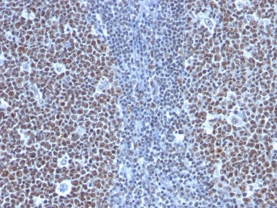
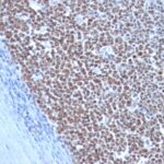
Cyclin B1 Previously Observed Antibody Staining Patterns
Observed Subcellular, Organelle Specific Staining Data:
Anti-CCNB1 antibody staining is expected to be primarily localized to the cytosol. There is variability in either the signal strength or the localization of signal in cytosol from cell to cell.
Observed Antibody Staining Data By Tissue Type:
Variations in Cyclin B1 antibody staining intensity in immunohistochemistry on tissue sections are present across different anatomical locations. An intense signal was observed in cells in the seminiferous ducts in testis, germinal center cells in the lymph node and tonsil and hematopoietic cells in the bone marrow. More moderate antibody staining intensity was present in cells in the seminiferous ducts in testis, germinal center cells in the lymph node and tonsil and hematopoietic cells in the bone marrow. Low, but measureable presence of Cyclin B1 could be seen in cells in the endometrial stroma in endometrium, cells in the tubules in kidney, glandular cells in the cervix, uterine and endometrium, Langerhans in skin, lymphoid tissue in appendix, melanocytes in skin, squamous epithelial cells in the cervix and uterine. We were unable to detect Cyclin B1 in other tissues. Disease states, inflammation, and other physiological changes can have a substantial impact on antibody staining patterns. These measurements were all taken in tissues deemed normal or from patients without known disease.
Observed Antibody Staining Data By Tissue Disease Status:
Tissues from cancer patients, for instance, have their own distinct pattern of Cyclin B1 expression as measured by anti-Cyclin B1 antibody immunohistochemical staining. The average level of expression by tumor is summarized in the table below. The variability row represents patient to patient variability in IHC staining.
| Sample Type | breast cancer | carcinoid | cervical cancer | colorectal cancer | endometrial cancer | glioma | head and neck cancer | liver cancer | lung cancer | lymphoma | melanoma | ovarian cancer | pancreatic cancer | prostate cancer | renal cancer | skin cancer | stomach cancer | testicular cancer | thyroid cancer | urothelial cancer |
|---|---|---|---|---|---|---|---|---|---|---|---|---|---|---|---|---|---|---|---|---|
| Signal Intensity | ++ | ++ | ++ | ++ | ++ | + | ++ | ++ | ++ | ++ | ++ | ++ | ++ | ++ | ++ | ++ | ++ | ++ | ++ | ++ |
| CCNB1 Variability | ++ | ++ | + | ++ | + | ++ | ++ | ++ | ++ | + | + | ++ | + | + | ++ | + | ++ | ++ | ++ | + |
| Cyclin B1 General Information | |
|---|---|
| Alternate Names | |
| G2/mitotic-specific cyclin-B1, CCNB1 | |
| Molecular Weight | |
| 55-62kDa | |
| Chromosomal Location | |
| 5q13.2 | |
| Curated Database and Bioinformatic Data | |
| Gene Symbol | CCNB1 |
| Entrez Gene ID | 891 |
| Ensemble Gene ID | ENSG00000134057 |
| RefSeq Protein Accession(s) | NP_001341773, NP_001341774, NP_114172 |
| RefSeq mRNA Accession(s) | NM_001354844, NM_001354845, NM_031966 |
| RefSeq Genomic Accession(s) | NC_000005, NC_018916, |
| UniProt ID(s) | P14635 |
| UniGene ID(s) | P14635 |
| HGNC ID(s) | 1579 |
| Cosmic ID(s) | CCNB1 |
| KEGG Gene ID(s) | hsa:891 |
| PharmGKB ID(s) | PA95 |
| General Description of Cyclin B1. | |
| It recognizes a protein of 55-62kDa, identified as cyclin B1. In mammals, cyclin B associates with inactive p34cdc2, which facilitates phosphorylation of p34cdc2 at aa 14Thr, 15Tyr. This maintains the inactive state until the end of G2-phase. The inactive cyclin B-p34cdc2 complex continues to accumulate in the cytoplasm until the completion of DNA synthesis, when Cdc25, a specific protein phosphatase, dephosphorylates aa 14Thr, 15Tyr of p34cdc2 rendering the complex active at the G2/M boundary. This mitotic kinase complex remains active until the metaphase/anaphase transition when cyclin B is degraded. This degradation process is ubiquitin-dependent, is necessary for the cell to exit mitosis. So, cyclin B-p34cdc2 plays a critical role in G2 to M transition. | |

Reviews
There are no reviews yet.