


 PDF Datasheet
Human, Mouse, and Rat Anti-NSE gamma Antibody Product Attributes
NSE gamma Previously Observed Antibody Staining Patterns
Observed Subcellular, Organelle Specific Staining Data:
Anti-ENO2 antibody staining is expected to be primarily localized to the cytosol, nucleus and plasma membrane. There is variability in either the signal strength or the localization of signal in cytosol and nucleus from cell to cell.
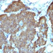
Observed Antibody Staining Data By Tissue Type:
Variations in NSE gamma antibody staining intensity in immunohistochemistry on tissue sections are present across different anatomical locations. An intense signal was observed in cells in the granular layer in cerebellum, neuronal cells in the caudate nucleus, cerebral cortex and hippocampus and neuropil in cerebral cortex. More moderate antibody staining intensity was present in cells in the granular layer in cerebellum, neuronal cells in the caudate nucleus, cerebral cortex and hippocampus and neuropil in cerebral cortex. Low, but measureable presence of NSE gamma could be seen inglial cells in the cerebral cortex and peripheral nerve in mesenchymal tissue. We were unable to detect NSE gamma in other tissues. Disease states, inflammation, and other physiological changes can have a substantial impact on antibody staining patterns. These measurements were all taken in tissues deemed normal or from patients without known disease.
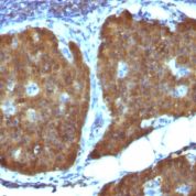
Observed Antibody Staining Data By Tissue Disease Status:
Tissues from cancer patients, for instance, have their own distinct pattern of NSE gamma expression as measured by anti-NSE gamma antibody immunohistochemical staining. The average level of expression by tumor is summarized in the table below. The variability row represents patient to patient variability in IHC staining.
| Sample Type | breast cancer | carcinoid | cervical cancer | colorectal cancer | endometrial cancer | glioma | head and neck cancer | liver cancer | lung cancer | lymphoma | melanoma | ovarian cancer | pancreatic cancer | prostate cancer | renal cancer | skin cancer | stomach cancer | testicular cancer | thyroid cancer | urothelial cancer |
|---|---|---|---|---|---|---|---|---|---|---|---|---|---|---|---|---|---|---|---|---|
| Signal Intensity | – | ++ | – | – | – | – | – | – | – | – | – | – | – | – | – | – | – | + | – | – |
| ENO2 Variability | + | + | + | + | + | ++ | + | + | + | + | + | + | + | + | ++ | + | + | +++ | ++ | + |
| NSE gamma General Information | |
|---|---|
| Alternate Names | |
| Gamma-enolase enolase 2, ENO2, neuron specific enolase, NSE, | |
| Molecular Weight | |
| 50kDa | |
| Chromosomal Location | |
| 12p13 | |
| Curated Database and Bioinformatic Data | |
| Gene Symbol | ENO2 |
| Entrez Gene ID | 2026 |
| Ensemble Gene ID | ENSG00000111674 |
| RefSeq Protein Accession(s) | NP_001966 |
| RefSeq mRNA Accession(s) | NM_001975 |
| RefSeq Genomic Accession(s) | NC_000012, NC_018923 |
| UniProt ID(s) | P09104, Q6FHV6 |
| UniGene ID(s) | P09104, Q6FHV6 |
| HGNC ID(s) | 3353 |
| Cosmic ID(s) | ENO2 |
| KEGG Gene ID(s) | hsa:2026 |
| PharmGKB ID(s) | PA27788 |
| General Description of NSE gamma. | |
| Recognizes a protein of about 50kDa, which is identified as gamma-enolase. Three isoenzymes of enolases are identified, alpha, beta and gamma. Alpha-isoform is expressed in most tissues, whereas beta-form is expressed predominantly in muscle tissue whereas gamma-enolase is found only in nervous tissue. These isoforms exist as both homodimers and heterodimers, and they play a role in converting phosphoglyceric acid to phosphenolpyruvic acid in the glycolytic pathway. NSE-gamma is a useful marker to identify peripheral nerves and tumors of neuro-endocrine origins, such as pheochromocytomas. It it be usually employed in combination with other markers such as Synaptophysin, Chromogranin A, and Neurofilament. | |
There are no reviews yet.