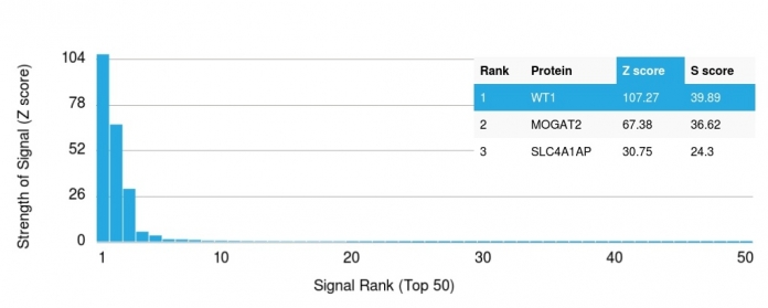
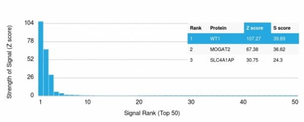
Analysis of Protein Array containing more than 19,000 full-length human proteins using Wilm’s Tumor Mouse Recombinant Monoclonal Antibody (rWT1/857). Z- and S- Score: The Z-score represents the strength of a signal that a monoclonal antibody (MAb) (in combination with a fluorescently-tagged anti-IgG secondary antibody) produces when binding to a particular protein on the HuProtTM array. Z-scores are described in units of standard deviations (SD’s) above the mean value of all signals generated on that array. If targets on HuProtTM are arranged in descending order of the Z-score, the S-score is the difference (also in units of SD’s) between the Z-score. S-score therefore represents the relative target specificity of a MAb to its intended target.  A MAb is considered to specific to its intended target, if the MAb has an S-score of at least 2.5. For example, if a MAb binds to protein X with a Z-score of 43 and to protein Y with a Z-score of 14, then the S-score for the binding of that MAb to protein X is equal to 29.
Analysis of Protein Array containing more than 19,000 full-length human proteins using Wilm’s Tumor Mouse Recombinant Monoclonal Antibody (rWT1/857). Z- and S- Score: The Z-score represents the strength of a signal that a monoclonal antibody (MAb) (in combination with a fluorescently-tagged anti-IgG secondary antibody) produces when binding to a particular protein on the HuProtTM array. Z-scores are described in units of standard deviations (SD’s) above the mean value of all signals generated on that array. If targets on HuProtTM are arranged in descending order of the Z-score, the S-score is the difference (also in units of SD’s) between the Z-score. S-score therefore represents the relative target specificity of a MAb to its intended target.  A MAb is considered to specific to its intended target, if the MAb has an S-score of at least 2.5. For example, if a MAb binds to protein X with a Z-score of 43 and to protein Y with a Z-score of 14, then the S-score for the binding of that MAb to protein X is equal to 29.SDS-PAGE Analysis of Purified Wilm’s Tumor Mouse Recombinant Monoclonal Antibody (rWT1/857).

Anti-Wilm s Tumor 1 Antibody Clone rWT1/857

$ 199.00$ 429.00
Please Select Product Options Below To View The Catalog Number.

SKU: 7490-MSM6
Species: Human
Tested Applications: IHC
Available Conjugates: Unconjugated
Isotype: Mouse IgG1, kappa
Mass Spec Validated?: Not MS Validated

Product NumberDescriptionPrice
7490-MSM6-P0 Size: 20 ug, Tag: Unconjugated $ 228.00
7490-MSM6-P1 Size: 100 ug, Tag: Unconjugated $ 488.00
7490-MSM6-P1ABX Size: 100 ug, Tag: Unconjugated $ 488.00
Shipping Information
In Stock.
Flat Rate Shipping Anywhere in the US: $45
Datasheets and Documentation
Product Datasheet
Certificate of Analysis and Tags (Coming Soon)
Lot Number:

Expiration Date:

Concentration (Write Lyophilized if Lyophilized):

Reconsitution Instructions (Leave Blank if Liquid):

Manufacture Date:

Purity:

Bioactivity (test results eg. IU/ml):


SKU: 7490-MSM6-P0
Wilm's Tumor 1 (WT1) (Wilm's Tumor & Mesothelial Marker) General Information
Alternate Names
WT1; AWT1; FWT1; GUD; NPHS4; WAGR; Wilms tumor 1
Molecular Weight
47-55kDa
Chromosomal Location
Ships on blue ice.
Curated Database and Bioinformatic Data
Gene Symbol7490
Entrez Gene IDWT1
UniProt ID(s)P19544
UniGene ID(s)Hs591980
COSMIC ID Link(s)WT1
KEGG Gene ID(s)hsa:7490
General Description of Wilm's Tumor 1 (WT1) (Wilm's Tumor & Mesothelial Marker).
Recognizes a 47-55kDa-tumor suppressor protein, identified as Wilm's Tumor (WT1) protein. The antibody reacts with all isoforms of the full-length WT1 and also identifies WT1 lacking exon 2-encoded amino acids, frequently found in subsets of sporadic Wilm s tumors. WT1, a sporadic and familial pediatric kidney tumor, is genetically heterogeneous. Wilm s tumor is associated with mutations of WT1, a zinc-finger transcription factor that is essential for the development of the metanephric kidney and the urogenital system. The WT1 gene is normally expressed in fetal kidney and mesothelium, and its expression has been suggested as a marker for Wilm s tumor and mesothelioma. WT1 protein has been identified in proliferative mesothelial cells, malignant mesothelioma, ovarian carcinoma, gonadoblastoma, nephroblastoma, and desmoplastic small round cell tumor. Lung adenocarcinomas rarely stain positive with this antibody. WT1 protein expression in mesothelial cells has become a reliable marker for the diagnosis of mesotheliomas.

Human Anti-Wilm's Tumor 1 (WT1) (Wilm's Tumor & Mesothelial Marker) Antibody Product Attributes

Species: Human
Tested Applications: IHC.
Clonality: Monoclonal
Anti-Wilm's Tumor 1 (WT1) (Wilm's Tumor & Mesothelial Marker) Antibody Clone: rWT1/857
Clone rWT1/857 Host and Isotype: Mouse IgG1, kappa
Anti-Human Wilm's Tumor 1 (WT1) (Wilm's Tumor & Mesothelial Marker) Positive Control Sample: K562 cells. Wilm s Tumor, mesothelioma or fetal kidney.
Cellular Localization of Antibody rWT1/857 Staining: Cytoplasmic
Buffer and Stabilizer: 10mM PBS with 0.05% BSA & 0.05% azide. Also available without BSA & Azide.
Antibody Concentration: 200 ug/ml
Antibody Purification Method:Protein A/G purified from Bioreactor concentrate.
Immunogen: Recombinant full-length human WT1 protein
Storage Conditions: Store at 2 to 8 C (refrigerate). Stable for 24 months when properly stored.

Wilm's Tumor 1 (WT1) (Wilm's Tumor & Mesothelial Marker) Previously Observed Antibody Staining Patterns

Observed Subcellular, Organelle Specific Staining Data:

Anti-WT1 antibody staining is expected to be primarily localized to the nucleoplasm.

Observed Antibody Staining Data By Tissue Type:

Variations in Wilm's Tumor 1 antibody staining intensity in immunohistochemistry on tissue sections are present across different anatomical locations. An intense signal was observed in myoepithelial cells in the breast, cells in the endometrial stroma in endometrium, glandular cells in the fallopian tube, cells in the glomeruli in kidney and cells in the seminiferous ducts in testis. More moderate antibody staining intensity was present in myoepithelial cells in the breast, cells in the endometrial stroma in endometrium, glandular cells in the fallopian tube, cells in the glomeruli in kidney and cells in the seminiferous ducts in testis. Low, but measureable presence of Wilm's Tumor 1 could be seen inglandular cells in the breast and epididymis, cells in the tubules in kidney, decidual cells in the placenta, trophoblastic cells in the placenta, glandular cells in the prostate and seminal vesicle, adipocytes in mesenchymal tissue, fibroblasts in mesenchymal tissue and cells in the red pulp in spleen. We were unable to detect Wilm's Tumor 1 in other tissues. Disease states, inflammation, and other physiological changes can have a substantial impact on antibody staining patterns. These measurements were all taken in tissues deemed normal or from patients without known disease.

Observed Antibody Staining Data By Tissue Disease Status:

Tissues from cancer patients, for instance, have their own distinct pattern of Wilm's Tumor 1 expression as measured by anti-Wilm's Tumor 1 antibody immunohistochemical staining. The average level of expression by tumor is summarized in the table below. The variability row represents patient to patient variability in IHC staining.

Sample Type breast cancer carcinoid cervical cancer colorectal cancer endometrial cancer glioma head and neck cancer liver cancer lung cancer lymphoma melanoma ovarian cancer pancreatic cancer prostate cancer renal cancer skin cancer stomach cancer testicular cancer thyroid cancer urothelial cancer
Signal Intensity - - - - - +++ - - - - ++ +++ - - - - - - - -
WT1 Variability ++ + + + + ++ + + + + ++ ++ + + ++ + + + + ++

Limitations and Warranty

enQuire Bio's product, Wilm's Tumor 1 (WT1) (Wilm's Tumor & Mesothelial Marker) MonoSpecific Antibody, is available for Research Use Only (RUO-Only). This antibody is guaranteed to work for a period of two years when properly stored.

There are no reviews yet.

Be the first to review “Anti-Wilm s Tumor 1 Antibody Clone rWT1/857”

Your email address will not be published. Required fields are marked *

Share a Protocol (View Reviewed Protocols Below)

Protocol Title

Materials

Methods

Notes


  protocol submission gif
enQuire Bio, LLC   8420 S Continental Divide Rd, #202   Littleton, CO 80127
Chat: See lower right corner. | enquire@enquirebio.com | Fax: 1-720-897-3730
Customer Service Available M-F 8AM-6PM MST or 24/7 Via Email

Anti-Wilm s Tumor 1 Antibody Clone rWT1/857

https://enquirebio.com/wp-content/uploads/2018/06/enQuire-Bio-7490-MSM6-P0-anti-Wilms-Tumor-1-WT1-Wilms-Tumor-Mesothelial-Marker-antibody-1-300x120.jpeghttps://enquirebio.com/wp-content/uploads/2018/06/enQuire-Bio-7490-MSM6-P0-anti-Wilms-Tumor-1-WT1-Wilms-Tumor-Mesothelial-Marker-antibody-238x300.jpeg