
Human, Pig, Mouse, and Rat Anti-Fibronectin Antibody Product Attributes
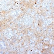
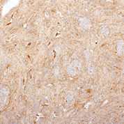
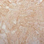
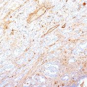
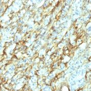
Fibronectin Previously Observed Antibody Staining Patterns
Observed Antibody Staining Data By Tissue Type:
Variations in Fibronectin antibody staining intensity in immunohistochemistry on tissue sections are present across different anatomical locations. An intense signal was observed in cells in the tubules in kidney. More moderate antibody staining intensity was present in cells in the tubules in kidney. Low, but measureable presence of Fibronectin could be seen inadipocytes in breast, cells in the endometrial stroma in endometrium, cells in the molecular layer in cerebellum, cells in the red pulp in spleen, cells in the seminiferous ducts in testis, cells in the white pulp in spleen, glandular cells in the appendix, breast, epididymis, rectum, salivary gland, seminal vesicle, small intestine, stomach and thyroid gland, glial cells in the caudate nucleus, hematopoietic cells in the bone marrow, hepatocytes in liver, macrophages in lung, neuronal cells in the hippocampus, peripheral nerve in mesenchymal tissue, respiratory epithelial cells in the bronchus and nasopharynx, squamous epithelial cells in the esophagus and oral mucosa, trophoblastic cells in the placenta and urothelial cells in the urinary bladder. We were unable to detect Fibronectin in other tissues. Disease states, inflammation, and other physiological changes can have a substantial impact on antibody staining patterns. These measurements were all taken in tissues deemed normal or from patients without known disease.
Observed Antibody Staining Data By Tissue Disease Status:
Tissues from cancer patients, for instance, have their own distinct pattern of Fibronectin expression as measured by anti-Fibronectin antibody immunohistochemical staining. The average level of expression by tumor is summarized in the table below. The variability row represents patient to patient variability in IHC staining.
| Sample Type | breast cancer | carcinoid | cervical cancer | colorectal cancer | endometrial cancer | glioma | head and neck cancer | liver cancer | lung cancer | lymphoma | melanoma | ovarian cancer | pancreatic cancer | prostate cancer | renal cancer | skin cancer | stomach cancer | testicular cancer | thyroid cancer | urothelial cancer |
|---|---|---|---|---|---|---|---|---|---|---|---|---|---|---|---|---|---|---|---|---|
| Signal Intensity | – | – | – | + | – | – | – | ++ | + | – | – | – | ++ | – | – | – | – | + | ++ | + |
| FN1 Variability | ++ | ++ | ++ | +++ | ++ | + | ++ | +++ | ++ | + | ++ | ++ | +++ | + | + | + | ++ | ++ | ++ | +++ |
| Fibronectin General Information | |
|---|---|
| Alternate Names | |
| Fibronectin, FN1 | |
| Molecular Weight | |
| 220kDa (monomer); 440kDa (dimer) | |
| Chromosomal Location | |
| 2q35 | |
| Curated Database and Bioinformatic Data | |
| Gene Symbol | FN1 |
| Entrez Gene ID | 2335 |
| Ensemble Gene ID | ENSG00000115414 |
| RefSeq Protein Accession(s) | XP_005246460, XP_005246464, XP_005246473, NP_997641, XP_005246461, XP_005246467, XP_005246469, XP_016859183, NP_001293060, NP_997639, NP_997647, XP_005246454, XP_005246466, XP_005246471, XP_016859182, NP_001293059, XP_005246458, XP_016859181, NP_001293058, XP_005246465, XP_005246468, NP_473375, XP_005246455, XP_005246459, NP_002017, NP_997643, XP_005246462, XP_005246456, XP_005246463, XP_016859184, NP_001293061 |
| RefSeq mRNA Accession(s) | XM_005246416, XM_017003692 NM_212476, XM_005246407, XM_005246410, XM_005246414, XM_017003694, NM_001306129, NM_001306130, NM_212474, XM_005246402, XM_005246408, XM_005246409, XM_005246399, NM_001306131, NM_054034, XM_005246403, XM_005246405, XM_005246406, XM_017003695, NM_002026, XM_005246398, XM_005246401, XM_005246404, XM_005246412, XM_017003693, NM_001306132, XM_005246397, XM_005246411, NM_212478, NM_212482, NM_212475 |
| RefSeq Genomic Accession(s) | NC_000002, NG_012196, NC_018913 |
| UniProt ID(s) | Q6N084, Q9UQS6, B7ZLE5, Q6MZF4, Q6MZM7, P02751 |
| UniGene ID(s) | Q6N084, Q9UQS6, B7ZLE5, Q6MZF4, Q6MZM7, P02751 |
| HGNC ID(s) | 3778 |
| Cosmic ID(s) | FN1 |
| KEGG Gene ID(s) | hsa:2335 |
| PharmGKB ID(s) | PA28194 |
| General Description of Fibronectin. | |
| Fibronectin is a soluble dimeric glycoprotein of 440kDa, which is present in cells, extracellular matrix,, blood. This MAb reacts with the cellular as well as plasma form of fibronectin. Reportedly, after iv administration, this MAb localizes to tumor vessels where it binds to the underlying basement. Epitope recognized by this antibody is not accessible in normal tissues to the circulating MAb indicating that it can be used to specifically target tumor vessels in vivo. TV-1 is reportedly useful for delivering vasoactive agents to tumors to induce increased vascular permeability or blood flow prior to treatment with chemotherapeutic drugs or MAbs. | |


There are no reviews yet.