
.jpg)
 PDF Datasheet
Human Anti-c-Myc Oncoprotein Antibody Product Attributes
c-Myc Oncoprotein Previously Observed Antibody Staining Patterns
Observed Subcellular, Organelle Specific Staining Data:
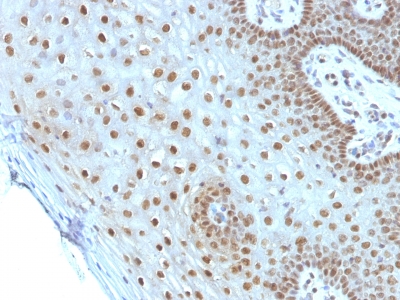
Anti-MYC antibody staining is expected to be primarily localized to the nucleoplasm.
Observed Antibody Staining Data By Tissue Type:
Variations in c-Myc Oncoprotein antibody staining intensity in immunohistochemistry on tissue sections are present across different anatomical locations. An intense signal was observed in neuronal cells in the caudate nucleus, Purkinje cells in the cerebellum, squamous epithelial cells in the cervix, uterine, neuronal cells in the hippocampus, pneumocytes in lung and cells in the seminiferous ducts in testis. More moderate antibody staining intensity was present in neuronal cells in the caudate nucleus, Purkinje cells in the cerebellum, squamous epithelial cells in the cervix, uterine, neuronal cells in the hippocampus, pneumocytes in lung and cells in the seminiferous ducts in testis. Low, but measureable presence of c-Myc Oncoprotein could be seen inhematopoietic cells in the bone marrow, glandular cells in the cervix, uterine, endothelial cells in the colon, glandular cells in the colon, endometrium and fallopian tube, glial cells in the hippocampus, bile duct cells in the liver, germinal center cells in the lymph node, ovarian stroma cells in the ovary, glandular cells in the parathyroid gland, trophoblastic cells in the placenta, glandular cells in the salivary gland, fibroblasts in skin, Langerhans in skin, melanocytes in skin, smooth muscle cells in the smooth muscle, adipocytes in mesenchymal tissue, fibroblasts in mesenchymal tissue, peripheral nerve in mesenchymal tissue, cells in the white pulp in spleen and glandular cells in the thyroid gland. We were unable to detect c-Myc Oncoprotein in other tissues. Disease states, inflammation, and other physiological changes can have a substantial impact on antibody staining patterns. These measurements were all taken in tissues deemed normal or from patients without known disease.
Observed Antibody Staining Data By Tissue Disease Status:
Tissues from cancer patients, for instance, have their own distinct pattern of c-Myc Oncoprotein expression as measured by anti-c-Myc Oncoprotein antibody immunohistochemical staining. The average level of expression by tumor is summarized in the table below. The variability row represents patient to patient variability in IHC staining.
| Sample Type | breast cancer | carcinoid | cervical cancer | colorectal cancer | endometrial cancer | glioma | head and neck cancer | liver cancer | lung cancer | lymphoma | melanoma | ovarian cancer | pancreatic cancer | prostate cancer | renal cancer | skin cancer | stomach cancer | testicular cancer | thyroid cancer | urothelial cancer |
|---|---|---|---|---|---|---|---|---|---|---|---|---|---|---|---|---|---|---|---|---|
| Signal Intensity | ++ | – | ++ | ++ | + | – | – | ++ | ++ | – | + | ++ | ++ | ++ | ++ | + | ++ | + | – | ++ |
| MYC Variability | ++ | ++ | ++ | ++ | + | ++ | ++ | + | ++ | ++ | +++ | ++ | ++ | +++ | ++ | ++ | ++ | ++ | ++ | + |
| c-Myc Oncoprotein General Information | |
|---|---|
| Alternate Names | |
| Myc, c-Myc, cMyc | |
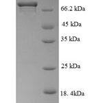
| Molecular Weight | |
| 62-64kDa | |
| Chromosomal Location | |
| 8q24.21 | |
| Curated Database and Bioinformatic Data | |
| Gene Symbol | MYC |
| Entrez Gene ID | 4609 |
| Ensemble Gene ID | ENSG00000136997 |
| RefSeq Protein Accession(s) | NP_001341799, NP_002458 |
| RefSeq mRNA Accession(s) | NM_001354870, NM_002467 |
| RefSeq Genomic Accession(s) | NC_000008, NG_007161, NC_018919 |
| UniProt ID(s) | P01106, A0A087WVR4 |
| UniGene ID(s) | P01106, A0A087WVR4 |
| HGNC ID(s) | 7553 |
| Cosmic ID(s) | MYC |
| KEGG Gene ID(s) | hsa:4609 |
| PharmGKB ID(s) | PA31353 |
| General Description of c-Myc Oncoprotein. | |
| The c-Myc protein is a transcription factor, which is encoded by the c-Myc gene on human chromosome 8q24. c-Myc is commonly activated in a variety of tumor cells, plays an important role in cellular proliferation, differentiation, apoptosis, cell cycle progression. The phosphorylation of c-Myc has been investigated, previous studies have suggested a functional association between phosphorylation at Thr58/Ser62 by glycogen synthase kinase 3, cyclin dependent kinase, ERK2, C-Jun N terminal Kinase (JNK) in cell proliferation, cell cycle regulation. Studies also have shown that c-Myc is essential for tumor cell development in vasculogenesis, angiogenesis that distribute blood throughout the cells,, which brought extensive attention in the development of new therapeutic approach for cancer treatment. | |
-150x150.jpg)

-150x150.jpg)
-150x150.jpg)
Reviews
There are no reviews yet.