
 PDF Datasheet
Human Anti-Calprotectin Antibody Product Attributes
Calprotectin Previously Observed Antibody Staining Patterns
Observed Subcellular, Organelle Specific Staining Data:
Anti-S100A9 antibody staining is expected to be primarily localized to the cell junctions and cytosol.
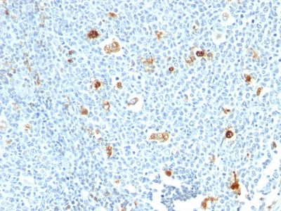
Observed Antibody Staining Data By Tissue Type:
Variations in Calprotectin antibody staining intensity in immunohistochemistry on tissue sections are present across different anatomical locations. An intense signal was observed in hematopoietic cells in the bone marrow, squamous epithelial cells in the cervix, uterine, esophagus and oral mucosa, epidermal cells in the skin, cells in the red pulp in spleen and squamous epithelial cells in the tonsil and vagina. More moderate antibody staining intensity was present in hematopoietic cells in the bone marrow, squamous epithelial cells in the cervix, uterine, esophagus and oral mucosa, epidermal cells in the skin, cells in the red pulp in spleen and squamous epithelial cells in the tonsil and vagina. Low, but measureable presence of Calprotectin could be seen in cells in the white pulp in spleen. We were unable to detect Calprotectin in other tissues. Disease states, inflammation, and other physiological changes can have a substantial impact on antibody staining patterns. These measurements were all taken in tissues deemed normal or from patients without known disease.
Observed Antibody Staining Data By Tissue Disease Status:
Tissues from cancer patients, for instance, have their own distinct pattern of Calprotectin expression as measured by anti-Calprotectin antibody immunohistochemical staining. The average level of expression by tumor is summarized in the table below. The variability row represents patient to patient variability in IHC staining.
| Sample Type | breast cancer | carcinoid | cervical cancer | colorectal cancer | endometrial cancer | glioma | head and neck cancer | liver cancer | lung cancer | lymphoma | melanoma | ovarian cancer | pancreatic cancer | prostate cancer | renal cancer | skin cancer | stomach cancer | testicular cancer | thyroid cancer | urothelial cancer |
|---|---|---|---|---|---|---|---|---|---|---|---|---|---|---|---|---|---|---|---|---|
| Signal Intensity | – | – | + | – | + | – | + | – | ++ | – | – | – | – | – | – | ++ | – | – | – | – |
| S100A9 Variability | ++ | + | ++ | + | ++ | + | ++ | ++ | ++ | ++ | + | ++ | ++ | ++ | + | ++ | + | + | + | ++ |
| Calprotectin General Information | |
|---|---|
| Alternate Names | |
| S100 calcium-binding protein A9, S100A9, migration inhibitory factor-related protein 14, MRP14, calgranulin B, S100A9 | |
| Molecular Weight | |
| 14kDa | |
| Chromosomal Location | |
| 1q21 | |
| Curated Database and Bioinformatic Data | |
| Gene Symbol | S100A9 |
| Entrez Gene ID | 6280 |
| Ensemble Gene ID | ENSG00000163220 |
| RefSeq Protein Accession(s) | NP_002956 |
| RefSeq mRNA Accession(s) | NM_002965 |
| RefSeq Genomic Accession(s) | NC_018912, NC_000001 |
| UniProt ID(s) | P06702 |
| UniGene ID(s) | P06702 |
| HGNC ID(s) | 10499 |
| Cosmic ID(s) | S100A9 |
| KEGG Gene ID(s) | hsa:6280 |
| PharmGKB ID(s) | PA34911 |
| General Description of Calprotectin. | |
| Recognizes the L1 or Calprotectin molecule, an intra-cytoplasmic antigen comprising of a 12kDa alpha chain, a 14kDa beta chain. Calprotectin comprises 60% of the cytoplasmic protein fraction of circulating polymorphonuclear granulocytes, is also found in monocytes, macrophages, ileal tissue eosinophils. Peripheral blood monocytes carry the antigen extra-, intracellularly, neutrophils only intracellularly. Calprotectin has antibacterial, antifungal, immunomodulating, antiproliferative effects. Besides this it is a potent chemotactic factor for neutrophils. Plasma concentrations are elevated in diseases associated with increased neutrophil activity, like inflammatory bowel disease. Granulocytes terminate their existence after transmigration through the intestinal wall. Therefore calprotectin is also detectable in feces. Elevated levels of calprotectin have been observed in body fluids such as plasma, saliva, gingival crevicular fluid, stools,, synovial fluid during infection, inflammatory conditions.This MAb reacts with neutrophils, monocytes, macrophages,, squamous mucosal epithelia, is important for identifying macrophages in tissue sections. | |
-150x150.jpg)
-150x150.jpg)
Reviews
There are no reviews yet.