
![Anti-CD25 Antibody [PC61.5] - Image 4](https://cdn-enquirebio.pressidium.com/wp-content/uploads/2017/10/enQuire-Bio-QAB34-B-100ug-anti-CD25-antibody-10.png)


 PDF Datasheet
Mouse Anti-CD25 Antibody Product Attributes
Species: Mouse
Tested Applications: Flow Cytometry.
Application Notes: See Product Datasheet for Recommended Dilution Range. Requires Experimental Optimization
Clonality: Monoclonal Antibody
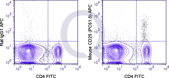
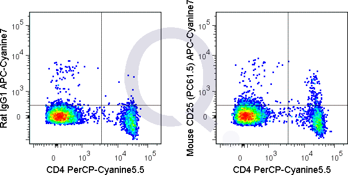
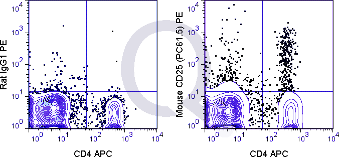
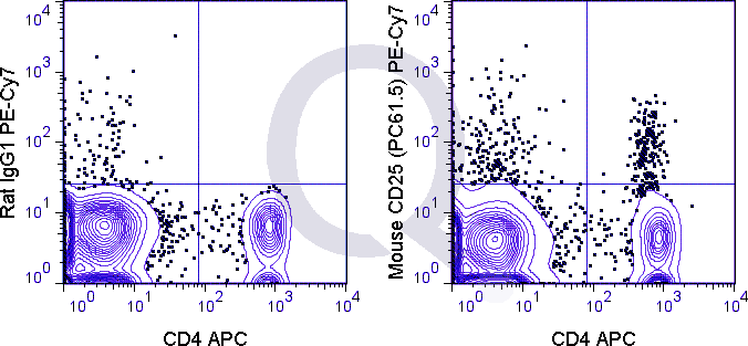
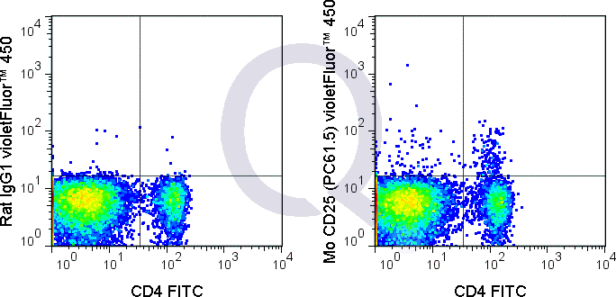
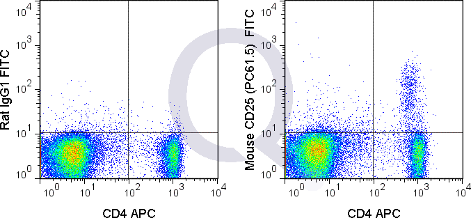
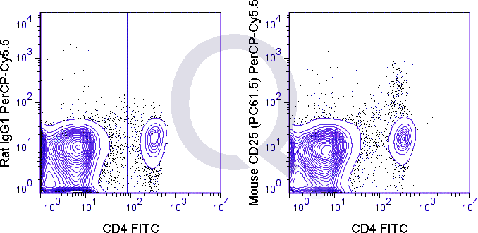
Anti-CD25 Antibody Clone: PC61.5
Clone PC61.5 Host and Isotype: Rat IgG1 lambda
Buffer and Stabilizer: 10 mM NaH2PO4, 150 mM NaCl, 0.09% NaN3, pH 7.2
Antibody Concentration: 0.5 mg/mL
Storage Conditions: 2-8C protected from light. Stable for 12 Months. Do Not Freeze.
CD25 Previously Observed Antibody Staining Patterns
Observed Antibody Staining Data By Tissue Type:
Variations in CD25 antibody staining intensity in immunohistochemistry on tissue sections are present across different anatomical locations. Low, but measureable presence of CD25 could be seen inhematopoietic cells in the bone marrow and lymphoid tissue in appendix. We were unable to detect CD25 in other tissues. Disease states, inflammation, and other physiological changes can have a substantial impact on antibody staining patterns. These measurements were all taken in tissues deemed normal or from patients without known disease.
| CD25 General Information | |
|---|---|
| Alternate Names | |
| Interleukin-2 receptor alpha, IL2RA, IL-2RA | |
| Curated Database and Bioinformatic Data | |
| Gene Symbol | Il2ra |
| Entrez Gene ID | 16184 |
| Ensemble Gene ID | ENSMUSG00000026770 |
| RefSeq Protein Accession(s) | NP_032393 |
| RefSeq mRNA Accession(s) | NM_008367 |
| RefSeq Genomic Accession(s) | NC_000068 |
| UniProt ID(s) | Q544I2, P01590 |
| UniGene ID(s) | Q544I2, P01590 |
| Cosmic ID(s) | Il2ra |
| KEGG Gene ID(s) | mmu:16184 |
| General Description of CD25. | |
| The PC61.5 antibody is specific for mouse CD25, a 55 kDa surface protein also known as the Interleukin-2 Receptor alpha chain, or IL-2R alpha. CD25 may bind IL-2 by itself, although with low affinity and without induction of cell signaling. CD25 is also expressed within a high-affinity complex, along with the IL-2R beta chain (CD122) and the common gamma chain (CD132), to form a signaling receptor complex. Expression of CD25 varies during developmental stages of T and B cells, is induced on activated mature T and B cells, and is present on subsets of dendritic cells. CD25 signaling as part of the IL-2 receptor complex triggers T cell activation and proliferation, as well as modulating the differentiation and function of Th17 cells, T regulatory (Treg) cells, and dendritic cells.The PC61.5 antibody is used as a marker for T cells, B cells and dendritic cell subsets. Expression of CD25, CD4 and the transcription factor Foxp3 is regarded as a phenotypic signature for Treg cells. As such, this antibody is widely used to distinguish Treg cells from nave or conventional T cells which are CD25-. This clone has also been reported for depletion of Treg cells in vivo (use format suitable for functional assays). | |
Selected References
Liang D, Zuo A, Shao H, Born WK, OBrian R, Kaplan HJ, and Sun D. 2012. J. Immunol. 188: 5785-5791. (in vivo blocking)Yu P, Steel JC, Zhang M, Morris JC, Waitz R, Fasso M, Allison JP, and Waldmann TA. 2012. Proc. Natl. Acad. Sci. 109:6187-6192. (in vivo Treg depletion)Billiard F, Lobry C, Darrasse-Jeze G, Waite J, Liu et al. 2012. Blood. 119: 4656-4664. (in vivo Treg depletion)Tang S, Moore ML, Grayson JM and Dubey P. 2012. Cancer Res. 72: 1975-1985. (in vivo Treg depletion)Lee L-F, Logronio K, Tu GH, Zhai W, Ni I, Mei L, Dilley J, Yu J, et al. 2012. Proc. Natl. Acad. Sci. 10.1073. (Flow cytometry).10F.9G2, J43, PC61 Koehn BH, Ford ML, Ferrer IR, Borom K, Gangappa S, Kirk AD, and Larsen CP. 2008. J. Immunol. 181:5313-5322. (in vivo blocking)Leithauser F, Meinhardt-Krajina T, Fink K, Wotschke B, Moller P and Reimann J. 2006. Am. J. Pathol. 168(6): 1898-1909. (Immunohistochemistry – frozen tissue)Hashimoto N, Nabholz M, MacDonald HR, and Zubler RH. 1986. Eur. J. Immunol. 16(3): 317-320. (Blocking)Ceredig R, Lowenthal JW, Nabholz M, and MacDonald R. 1985. Nature. 314:98-100 (Immunohistochemistry)Lowenthal JW, Zulber RH, Nabholz M, and MacDonald HR. 1985. Nature. 315(6021): 669-672. (Immunoprecipitation, Blocking)
Limitations and Warranty
enQuire Bio’s Mouse Anti-CD25 Monoclonal is available for Research Use Only. This antibody is guaranteed to work for a period of two years when properly stored.
There are no reviews yet.