

![Anti-Human/Mouse CD44 Antibody [IM7] - Image 4](https://cdn-enquirebio.pressidium.com/wp-content/uploads/2017/10/enQuire-Bio-QAB39-B-100ug-anti-CD44-HCAM-antibody-10.png)


 PDF Datasheet
Human and Mouse Anti-CD44 / HCAM Antibody Product Attributes
CD44 / HCAM Previously Observed Antibody Staining Patterns
Observed Subcellular, Organelle Specific Staining Data:
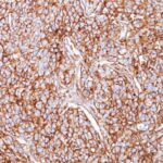
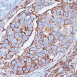
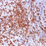
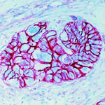
Staining with anti-CD44 / HCAM antibody reveals CD44 / HCAM expression is expected to be primarily localized to the golgi apparatus and plasma membrane.
Observed Antibody Staining Data By Tissue Type:
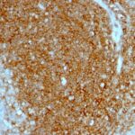
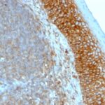
Variations in CD44 / HCAM antibody staining intensity in immunohistochemistry on tissue sections are present across different anatomical locations. An intense signal was observed in epidermal cells in the skin, glandular cells in the breast, cervix, uterine, prostate and salivary gland, hematopoietic cells in the bone marrow, keratinocytes in skin, Langerhans in skin, melanocytes in skin, respiratory epithelial cells in the bronchus and nasopharynx, squamous epithelial cells in the cervix, uterine, esophagus, oral mucosa, tonsil and vagina and urothelial cells in the urinary bladder. More moderate antibody staining intensity was present in epidermal cells in the skin, glandular cells in the breast, cervix, uterine, prostate and salivary gland, hematopoietic cells in the bone marrow, keratinocytes in skin, Langerhans in skin, melanocytes in skin, respiratory epithelial cells in the bronchus and nasopharynx, squamous epithelial cells in the cervix, uterine, esophagus, oral mucosa, tonsil and vagina and urothelial cells in the urinary bladder. Low, but measureable presence of CD44 / HCAM could be seen inadipocytes in mesenchymal tissue, cells in the glomeruli in kidney, cells in the tubules in kidney, decidual cells in the placenta, endothelial cells in the cerebral cortex and colon, follicle cells in the ovary, glandular cells in the fallopian tube, rectum and seminal vesicle, Leydig cells in the testis, neuronal cells in the cerebral cortex, neuropil in cerebral cortex, pneumocytes in lung and trophoblastic cells in the placenta. We were unable to detect CD44 / HCAM in other tissues. Disease states, inflammation, and other physiological changes can have a substantial impact on antibody staining patterns. These measurements were all taken in tissues deemed normal or from patients without known disease.
Observed Antibody Staining Data By Tissue Disease Status:
Tissues from cancer patients, for instance, have their own distinct pattern of CD44 / HCAM expression as measured by anti-CD44 / HCAM antibody immunohistochemical staining. The average level of expression by tumor is summarized in the table below. The variability row represents patient to patient variability in IHC staining.
| Sample Type | breast cancer | carcinoid | cervical cancer | colorectal cancer | endometrial cancer | glioma | head and neck cancer | liver cancer | lung cancer | lymphoma | melanoma | ovarian cancer | pancreatic cancer | prostate cancer | renal cancer | skin cancer | stomach cancer | testicular cancer | thyroid cancer | urothelial cancer |
|---|---|---|---|---|---|---|---|---|---|---|---|---|---|---|---|---|---|---|---|---|
| Signal Intensity | ++ | – | +++ | ++ | ++ | +++ | +++ | + | + | +++ | +++ | + | ++ | ++ | – | ++ | – | – | ++ | – |
| CD44 Variability | ++ | + | + | ++ | ++ | ++ | + | ++ | ++ | ++ | + | ++ | ++ | ++ | ++ | ++ | ++ | ++ | ++ | ++ |
| CD44 / HCAM General Information | |
|---|---|
| Alternate Names | |
| CD44, HCAM, CDW44, HUTCH-I, IN, LHR, CSPG8, MC56, MDU2, MDU3, ECMR-III, MIC4, Pgp1, HCELL | |
| Curated Database and Bioinformatic Data | |
| Gene Symbol | Cd44 |
| Entrez Gene ID | 12505 |
| Ensemble Gene ID | ENSMUSG00000005087 |
| RefSeq Protein Accession(s) | XP_006498711, NP_001171256, NP_001171257, XP_006498709, NP_033981, XP_006498708, XP_006498712, NP_001034240, NP_001171258, XP_006498710, NP_001034239 |
| RefSeq mRNA Accession(s) | XM_006498649, NM_009851, XM_006498647, XM_006498645, XM_006498646, NM_001039150, NM_001177787, NM_001039151, XM_006498648, NM_001177785, NM_001177786 |
| RefSeq Genomic Accession(s) | NC_000068 |
| UniProt ID(s) | P15379, A2APM1, Q3U8S1, A2APM2, Q80X37, A2APM4, A2APM3 |
| UniGene ID(s) | P15379, A2APM1, Q3U8S1, A2APM2, Q80X37, A2APM4, A2APM3 |
| Cosmic ID(s) | Cd44 |
| KEGG Gene ID(s) | mmu:12505 |
| General Description of CD44 / HCAM. | |
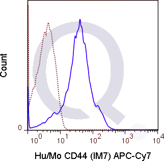
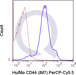
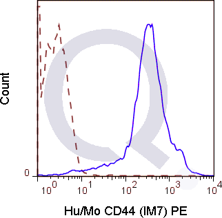
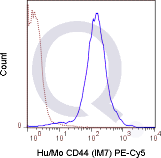
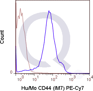
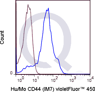
| The IM7 antibody recognizes CD44, a ubiquitously expressed cell surface receptor which is important for extracellular matrix organization, cell-cell and cell-matrix adhesion and migration. CD44 may be expressed in a number of different isoforms (splice variants) from the most typical or standard form, known as CD44s, to variants designated CD44v, e.g. CD44v1 or CD44v6. These receptors interact with several ligands, but most often associate with an extracellular matrix component hyaluronate, through which it mediates adhesion.The IM7 antibody may be used for detection of all isoforms of CD44, as it recognizes constant epitopes near the extracellular proximal domain. (Xu et al, 2002, J. Leukoc. Biol. 72:1133-1141). It has been reported to be cross-reactive with many non-human species including Baboon, Chimpanzee, Cynomolgus, Rhesus, Horse, Cow, Pig, Canine and Cat CD44. | |
Selected References
Limitations and Warranty
| Size | |
|---|---|
| Tag | APC, APC-Cy7, Biotin, FITC, PE, PE-Cy5, PE-Cy7, PerCP-Cy5.5, Qfluor™ 630, Qfluor™ 710, Unconjugated, V450 |
| Buffer and Stabilizer | 10 mM NaH2PO4, 150 mM NaCl, 0.09% NaN3, 0.1, pH7.2, 10 mM NaH2PO4, 150 mM NaCl, 0.09% NaN3, 0.1% gelatin, pH7.2, 10 mM NaH2PO4, 150 mM NaCl, 0.09% NaN3, pH 7.2 |
| Product Type | |
| Host | |
| Isotype | |
| Applications | |
| Species | |
| Mass Spec Validated? |
Only logged in customers who have purchased this product may leave a review.
![Analysis of Mass Spec data (dashed-line) of fractions stained with CD44 / HCAM <a href="https://enquirebio.com/validation-project-details/" target="_blank">MS-QAVA™ monoclonal antibody</a> [Clone: HCAM/918] (solid-line), reveals that less than 12.6% of signal is attributable to non-specific binding of anti-CD44 / HCAM Std. Anti-Human, Primate [Clone HCAM/918] to targets other than CD44 protein. Even frequently cited antibodies have much greater non-specific interactions, averaging over 30%. Data in image is from analysis in Jurkat, U202 and HeLa cells.](https://cdn-enquirebio.pressidium.com/wp-content/uploads/2017/10/enQuire-Bio-960-MSM2-P1-anti-CD44-HCAM-antibody-150x150.png)
Reviews
There are no reviews yet.