
 PDF Datasheet
Human and Rat (-) Anti-Chromogranin A / CHGA Antibody Product Attributes
Chromogranin A / CHGA Previously Observed Antibody Staining Patterns
Observed Subcellular, Organelle Specific Staining Data:
Anti-CHGA antibody staining is expected to be primarily localized to the vesicles.
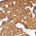
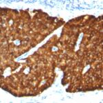
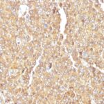
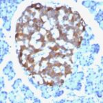
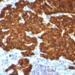
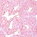
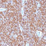
Observed Antibody Staining Data By Tissue Type:
Variations in Chromogranin A / CHGA antibody staining intensity in immunohistochemistry on tissue sections are present across different anatomical locations. An intense signal was observed in glandular cells in the parathyroid gland and islets of Langerhans in pancreas. More moderate antibody staining intensity was present in glandular cells in the parathyroid gland and islets of Langerhans in pancreas. Low, but measureable presence of Chromogranin A / CHGA could be seen inneuronal cells in the caudate nucleus, cerebral cortex and hippocampus, respiratory epithelial cells in the bronchus and neuronal cells in the caudate nucleus and cerebral cortex and hippocampus. We were unable to detect Chromogranin A / CHGA in other tissues. Disease states, inflammation, and other physiological changes can have a substantial impact on antibody staining patterns. These measurements were all taken in tissues deemed normal or from patients without known disease.
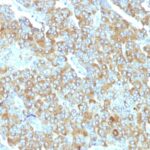
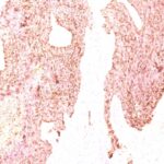
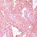
Observed Antibody Staining Data By Tissue Disease Status:
Tissues from cancer patients, for instance, have their own distinct pattern of Chromogranin A / CHGA expression as measured by anti-Chromogranin A / CHGA antibody immunohistochemical staining. The average level of expression by tumor is summarized in the table below. The variability row represents patient to patient variability in IHC staining.
| Sample Type | breast cancer | carcinoid | cervical cancer | colorectal cancer | endometrial cancer | glioma | head and neck cancer | liver cancer | lung cancer | lymphoma | melanoma | ovarian cancer | pancreatic cancer | prostate cancer | renal cancer | skin cancer | stomach cancer | testicular cancer | thyroid cancer | urothelial cancer |
|---|---|---|---|---|---|---|---|---|---|---|---|---|---|---|---|---|---|---|---|---|
| Signal Intensity | – | +++ | – | – | – | – | – | – | – | – | – | – | – | – | – | – | – | – | – | – |
| CHGA Variability | + | + | + | + | ++ | + | ++ | + | + | + | + | + | + | + | + | ++ | ++ | + | + | + |
| Chromogranin A / CHGA General Information | |
|---|---|
| Alternate Names | |
| TIMP metallopeptidase inhibitor 1, TIMP1, Tissue Inhibitor of Metalloproteinase 1 | |
| Molecular Weight | |
| 68-75kDa | |
| Chromosomal Location | |
| 14q32.12 | |
| Curated Database and Bioinformatic Data | |
| Gene Symbol | CHGA |
| Entrez Gene ID | 1113 |
| Ensemble Gene ID | ENSG00000276781, ENSG00000100604 |
| RefSeq Protein Accession(s) | NP_001288619, XP_011534672, NP_001266 |
| RefSeq mRNA Accession(s) | XM_011536370, NM_001301690, NM_001275 |
| RefSeq Genomic Accession(s) | NC_000014, NG_187601, NC_018925 |
| UniProt ID(s) | Q86T07, P10645, G5E968 |
| UniGene ID(s) | Q86T07, P10645, G5E968 |
| HGNC ID(s) | 1929 |
| Cosmic ID(s) | CHGA |
| KEGG Gene ID(s) | hsa:1113 |
| PharmGKB ID(s) | PA26461 |
| General Description of Chromogranin A / CHGA. | |
| Chromogranin A is present in neuroendocrine cells throughout the body, including the neuroendocrine cells of the large, small intestine, adrenal medulla, pancreatic islets. It is an excellent marker for carcinoid tumors, pheochromocytomas, paragangliomas,, other neuroendocrine tumors. Co-expression of chromogranin A, neuron specific enolase (NSE) is common in neuroendocrine neoplasms. Reportedly, co-expression of certain keratins, chromogranin indicates neuroendocrine lineage. The presence of strong anti-chromogranin staining, absence of anti-keratin staining should raise the possibility of paraganglioma. The co-expression of chromogranin, NSE is typical of neuroendocrine neoplasms. Most pituitary adenomas, prolactinomas readily express chromogranin. | |

Reviews
There are no reviews yet.