

 PDF Datasheet
Human, Rat, Cow, Goat, and Pig Anti-Cytokeratin 17 Antibody Product Attributes
Cytokeratin 17 Previously Observed Antibody Staining Patterns
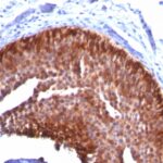
Observed Subcellular, Organelle Specific Staining Data:
Anti-krt17 antibody staining is expected to be primarily localized to the intermediate filaments. There is variability in either the signal strength or the localization of signal in intermediate filaments from cell to cell.
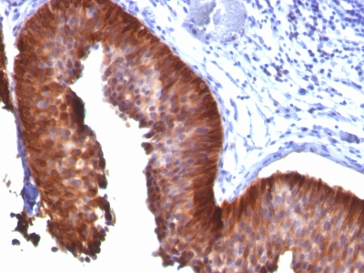
Observed Antibody Staining Data By Tissue Type:
Variations in Cytokeratin 17 antibody staining intensity in immunohistochemistry on tissue sections are present across different anatomical locations. An intense signal was observed in glandular cells in the prostate and salivary gland, myoepithelial cells in the breast and urothelial cells in the urinary bladder. More moderate antibody staining intensity was present in glandular cells in the prostate and salivary gland, myoepithelial cells in the breast and urothelial cells in the urinary bladder. Low, but measureable presence of Cytokeratin 17 could be seen inexocrine glandular cells in the pancreas, glandular cells in the fallopian tube and squamous epithelial cells in the esophagus and oral mucosa. We were unable to detect Cytokeratin 17 in other tissues. Disease states, inflammation, and other physiological changes can have a substantial impact on antibody staining patterns. These measurements were all taken in tissues deemed normal or from patients without known disease.
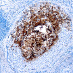
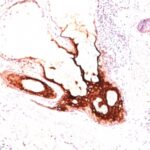
Observed Antibody Staining Data By Tissue Disease Status:
Tissues from cancer patients, for instance, have their own distinct pattern of Cytokeratin 17 expression as measured by anti-Cytokeratin 17 antibody immunohistochemical staining. The average level of expression by tumor is summarized in the table below. The variability row represents patient to patient variability in IHC staining.
| Sample Type | breast cancer | carcinoid | cervical cancer | colorectal cancer | endometrial cancer | glioma | head and neck cancer | liver cancer | lung cancer | lymphoma | melanoma | ovarian cancer | pancreatic cancer | prostate cancer | renal cancer | skin cancer | stomach cancer | testicular cancer | thyroid cancer | urothelial cancer |
|---|---|---|---|---|---|---|---|---|---|---|---|---|---|---|---|---|---|---|---|---|
| Signal Intensity | – | – | +++ | – | – | – | +++ | – | ++ | – | – | – | – | – | – | +++ | – | – | – | +++ |
| KRT17 Variability | + | + | + | + | ++ | + | ++ | ++ | ++ | + | + | ++ | ++ | + | + | ++ | + | + | + | ++ |
| Cytokeratin 17 General Information | |
|---|---|
| Alternate Names | |
| Keratin, type I cytoskeletal 17, cytokeratin 17, KRT17 | |
| Molecular Weight | |
| 46kDa | |
| Chromosomal Location | |
| 17q21.2 | |
| Curated Database and Bioinformatic Data | |
| Gene Symbol | KRT17 |
| Entrez Gene ID | 3872 |
| Ensemble Gene ID | ENSG00000128422 |
| RefSeq Protein Accession(s) | NP_000413 |
| RefSeq mRNA Accession(s) | NM_000422 |
| RefSeq Genomic Accession(s) | NC_018928, NG_008625, NG_009090, NC_000017 |
| UniProt ID(s) | Q14666, Q04695 |
| UniGene ID(s) | Q14666, Q04695 |
| HGNC ID(s) | 6427 |
| Cosmic ID(s) | KRT17 |
| KEGG Gene ID(s) | hsa:3872 |
| PharmGKB ID(s) | PA30214 |
| General Description of Cytokeratin 17. | |
| Cytokeratin 17 (CK17) is normally expressed in the basal cells of complex epithelia but not in stratified or simple epithelia. Antibody to CK17 is an excellent tool to distinguish myoepithelial cells from luminal epithelium of various glands such as mammary, sweat, salivary. CK17 is expressed in epithelial cells of various origins, such as bronchial epithelial cells, skin appendages. It may be considered as epithelial stem cell marker because CK17 Ab marks basal cell differentiation. CK17 is expressed in SCLC much higher than in LADC. Eighty-five percent of the triple negative breast carcinomas immunoreact with basal cytokeratins including anti-CK17. Also important is that cases of triple negative breast carcinoma with expression of CK17 show an aggressive clinical course. The histologic differentiation of ampullary cancer, intestinal vs. pancreatobiliary, is very important for treatment. Usually anti-CK17, anti-MUC1 immunoreactivity represents pancreatobiliary subtype whereas anti-MUC2, anti-CDX-2 positivity defines intestinal subtype. | |

There are no reviews yet.