
 PDF Datasheet
Human, Monkey, Cow, Canine, Rabbit, Mouse, Rat, and Chicken Anti-Cytokeratin Antibody Product Attributes
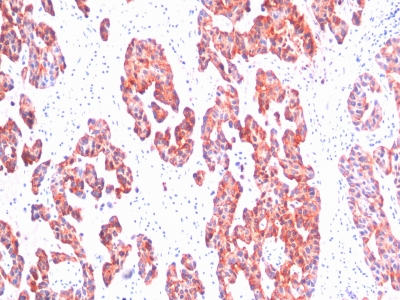
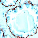
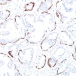
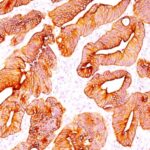
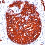
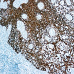
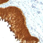
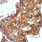
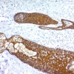
Cytokeratin Previously Observed Antibody Staining Patterns
Observed Antibody Staining Data By Tissue Type:
Variations in Cytokeratin antibody staining intensity in immunohistochemistry on tissue sections are present across different anatomical locations. Low, but measureable presence of Cytokeratin could be seen in. We were unable to detect Cytokeratin in other tissues. Disease states, inflammation, and other physiological changes can have a substantial impact on antibody staining patterns. These measurements were all taken in tissues deemed normal or from patients without known disease.
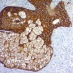
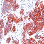
Observed Antibody Staining Data By Tissue Disease Status:
Tissues from cancer patients, for instance, have their own distinct pattern of Cytokeratin expression as measured by anti-Cytokeratin antibody immunohistochemical staining. The average level of expression by tumor is summarized in the table below. The variability row represents patient to patient variability in IHC staining.
| Sample Type | breast cancer | carcinoid | cervical cancer | colorectal cancer | endometrial cancer | glioma | head and neck cancer | liver cancer | lung cancer | lymphoma | melanoma | ovarian cancer | pancreatic cancer | prostate cancer | renal cancer | skin cancer | stomach cancer | testicular cancer | thyroid cancer | urothelial cancer |
|---|---|---|---|---|---|---|---|---|---|---|---|---|---|---|---|---|---|---|---|---|
| Signal Intensity | + | – | – | + | + | – | + | + | – | – | – | + | + | – | – | – | – | – | + | – |
| KRT76 Variability | ++ | ++ | ++ | ++ | ++ | + | ++ | ++ | ++ | + | ++ | ++ | ++ | ++ | ++ | + | ++ | ++ | ++ | ++ |
| Cytokeratin General Information | |
|---|---|
| Alternate Names | |
| KRT2B, KRT2P, HUMCYT2A, Keratin, type II Cytoskeletal 2 oral, K76, Keratin 2p (K2P), Keratin-76, Cytokeratin-2P (CK-2P, Type-II Keratin Kb9, anti-KRT2B antibody, anti-KRT2P antibody, anti-HUMCYT2A antibody, anti-Keratin antibody, anti-type II Cytoskeletal 2 oral antibody, anti-K76 antibody, anti-Keratin 2p (K2P) antibody, anti-Keratin-76 antibody, anti-Cytokeratin-2P (CK-2P antibody, anti-Type-II Keratin Kb9 antibody | |
| Molecular Weight | |
| 67kDa (CK1); 64kDa (CK3); 59kDa (CK4); 58kDa (CK5); 56kDa (CK6); 52kDa (CK8) | |
| Chromosomal Location | |
| 12q13.13 | |
| Curated Database and Bioinformatic Data | |
| Gene Symbol | KRT76 |
| Entrez Gene ID | 51350 |
| Ensemble Gene ID | ENSG00000185069 |
| RefSeq Protein Accession(s) | NP_056932 |
| RefSeq mRNA Accession(s) | NM_015848, |
| RefSeq Genomic Accession(s) | NG_012420, NC_000012, NC_018923 |
| UniProt ID(s) | Q01546 |
| UniGene ID(s) | Q01546 |
| HGNC ID(s) | 24430 |
| Cosmic ID(s) | KRT76 |
| KEGG Gene ID(s) | hsa:51350 |
| PharmGKB ID(s) | PA147357785 |
| General Description of Cytokeratin. | |
| This MAb recognizes basic (Type II or HMW) cytokeratins, which include 67kDa (CK1); 64kDa (CK3); 59kDa (CK4); 58kDa (CK5); 56kDa (CK6); 52kDa (CK8). Twenty human keratins are resolved with two-dimensional gel electrophoresis into acidic (pI 6.0) subfamilies. The acidic keratins have molecular weights (MW) of 56.5, 55, 51, 50, 50, 48, 46, 45,, 40kDa. This MAb recognizes the 65-67, 64, 59, 58, 56,, 52kDa keratins of basic subfamily. Many studies have shown the usefulness of keratins as markers in cancer research, tumor diagnosis. SPM115/SPM116 is a broad spectrum anti pan-keratin antibody cocktail, which differentiates epithelial tumors from non-epithelial tumors e.g. squamous vs. adenocarcinoma of the lung, liver carcinoma, breast cancer,, esophageal cancer. | |

Reviews
There are no reviews yet.