
.jpg)
.jpg)
 PDF Datasheet
Human and Rat Anti-Fascin-1 Antibody Product Attributes
Fascin-1 Previously Observed Antibody Staining Patterns
Observed Subcellular, Organelle Specific Staining Data:
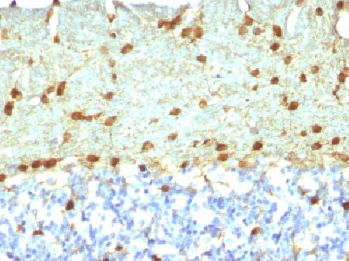
Variations in Fascin-1 antibody staining intensity in immunohistochemistry on tissue sections are present across different anatomical locations. An intense signal was observed in cells in the glomeruli in kidney, cells in the molecular layer in cerebellum, cells in the seminiferous ducts in testis, glandular cells in the breast, glial cells in the caudate nucleus and cerebral cortex, Langerhans in skin and squamous epithelial cells in the tonsil. More moderate antibody staining intensity was present in cells in the glomeruli in kidney, cells in the molecular layer in cerebellum, cells in the seminiferous ducts in testis, glandular cells in the breast, glial cells in the caudate nucleus and cerebral cortex, Langerhans in skin and squamous epithelial cells in the tonsil. Low, but measureable presence of Fascin-1 could be seen indecidual cells in the placenta, germinal center cells in the lymph node and tonsil, hematopoietic cells in the bone marrow, keratinocytes in skin, lymphoid tissue in appendix, neuronal cells in the caudate nucleus, neuropil in cerebral cortex, non-germinal center cells in the tonsil, pneumocytes in lung, squamous epithelial cells in the cervix and uterine, esophagus, oral mucosa and vagina. We were unable to detect Fascin-1 in other tissues. Disease states, inflammation, and other physiological changes can have a substantial impact on antibody staining patterns. These measurements were all taken in tissues deemed normal or from patients without known disease.
Observed Antibody Staining Data By Tissue Type:
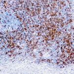
Tissues from cancer patients, for instance, have their own distinct pattern of Fascin-1 expression as measured by anti-Fascin-1 antibody immunohistochemical staining. The average level of expression by tumor is summarized in the table below. The variability row represents patient to patient variability in IHC staining.
| Sample Type | breast cancer | carcinoid | cervical cancer | colorectal cancer | endometrial cancer | glioma | head and neck cancer | liver cancer | lung cancer | lymphoma | melanoma | ovarian cancer | pancreatic cancer | prostate cancer | renal cancer | skin cancer | stomach cancer | testicular cancer | thyroid cancer | urothelial cancer |
|---|---|---|---|---|---|---|---|---|---|---|---|---|---|---|---|---|---|---|---|---|
| Signal Intensity | – | – | ++ | – | – | ++ | +++ | – | ++ | – | + | – | – | – | – | ++ | – | ++ | – | ++ |
| FSCN1 Variability | + | ++ | ++ | + | ++ | ++ | + | ++ | ++ | ++ | ++ | ++ | + | + | + | ++ | + | ++ | + | ++ |
| Fascin-1 General Information | |
|---|---|
| Alternate Names | |
| Fascin, FSCN1 | |
| Molecular Weight | |
| 55kDa | |
| Chromosomal Location | |
| 7p22.1 | |
| Curated Database and Bioinformatic Data | |
| Gene Symbol | FSCN1 |
| Entrez Gene ID | 6624 |
| Ensemble Gene ID | ENSG00000075618 |
| RefSeq Protein Accession(s) | NP_003079 |
| RefSeq mRNA Accession(s) | NM_003088 |
| RefSeq Genomic Accession(s) | NC_000007, NG_030004, NC_018918 |
| UniProt ID(s) | Q16658, B3KTA3 |
| UniGene ID(s) | Q16658, B3KTA3 |
| HGNC ID(s) | 11148 |
| Cosmic ID(s) | FSCN1 |
| KEGG Gene ID(s) | hsa:6624 |
| PharmGKB ID(s) | PA128394534 |
| General Description of Fascin-1. | |
| Recognizes a protein of 55kDa, which is identified as fascin-1. Its actin binding ability is regulated by phosphorylation. Antibody to fascin-1 is a very sensitive marker for Reed-Sternberg cells, variants in nodular sclerosis, mixed cellularity,, lymphocyte depletion Hodgkin s disease. It is uniformly negative in lymphoid cells, plasma cells,, myeloid cells. Fascin-1 is also expressed in dendritic cells. This marker may be helpful to distinguish between Hodgkin lymphoma, non-Hodgkin lymphoma in difficult cases. Also, the lack of expression of fascin-1 in the neoplastic follicles in follicular lymphoma may be helpful in distinguishing these lymphomas from reactive follicular hyperplasia in which the number of follicular dendritic cells is normal or increased. Antibody to fascin-1 has been suggested as a prognostic marker in neuroendocrine neoplasms of the lung as well as in ovarian cancer. Fascin-1 expression may be induced by Epstein-Barr virus (EBV) infection of B cells with the possibility that viral induction of fascin in lymphoid or other cell types must also be considered in EBV-positive cases. | |
-150x150.jpg)
-150x150.jpg)
-150x150.jpg)
Reviews
There are no reviews yet.