
![Anti-FCER1A Antibody [MAR-1]](https://cdn-enquirebio.pressidium.com/wp-content/uploads/2017/10/enQuire-Bio-QAB91-F-100ug-anti-FcERI-FCER1A-antibody-10.png)
![Anti-FCER1A Antibody [MAR-1] - Image 3](https://cdn-enquirebio.pressidium.com/wp-content/uploads/2017/10/enQuire-Bio-QAB91-PE-100ug-anti-FcERI-FCER1A-antibody-10.png)
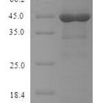
 PDF Datasheet
Mouse Anti-FcERI / FCER1A Antibody Product Attributes
FcERI / FCER1A Previously Observed Antibody Staining Patterns
Observed Antibody Staining Data By Tissue Type:
Variations in FcERI / FCER1A antibody staining intensity in immunohistochemistry on tissue sections are present across different anatomical locations. Low, but measureable presence of FcERI / FCER1A could be seen in cells in the red pulp in spleen, endothelial cells in the colon, epidermal cells in the skin, glandular cells in the adrenal gland, appendix, colon, duodenum, fallopian tube, rectum, seminal vesicle and thyroid gland, hematopoietic cells in the bone marrow, Leydig cells in the testis, melanocytes in skin, myocytes in heart muscle and skeletal muscle, myoepithelial cells in the breast, neuronal cells in the hippocampus, respiratory epithelial cells in the nasopharynx, trophoblastic cells in the placenta and urothelial cells in the urinary bladder. We were unable to detect FcERI / FCER1A in other tissues. Disease states, inflammation, and other physiological changes can have a substantial impact on antibody staining patterns. These measurements were all taken in tissues deemed normal or from patients without known disease.
Observed Antibody Staining Data By Tissue Disease Status:
Tissues from cancer patients, for instance, have their own distinct pattern of FcERI / FCER1A expression as measured by anti-FcERI / FCER1A antibody immunohistochemical staining. The average level of expression by tumor is summarized in the table below. The variability row represents patient to patient variability in IHC staining.
| Sample Type | breast cancer | carcinoid | cervical cancer | colorectal cancer | endometrial cancer | glioma | head and neck cancer | liver cancer | lung cancer | lymphoma | melanoma | ovarian cancer | pancreatic cancer | prostate cancer | renal cancer | skin cancer | stomach cancer | testicular cancer | thyroid cancer | urothelial cancer |
|---|---|---|---|---|---|---|---|---|---|---|---|---|---|---|---|---|---|---|---|---|
| Signal Intensity | – | – | – | – | – | – | – | + | – | – | – | – | – | – | – | – | – | – | – | – |
| FCER1A Variability | ++ | ++ | + | ++ | + | ++ | + | ++ | + | + | + | + | + | + | + | + | + | + | ++ | + |
| FcERI / FCER1A General Information | |
|---|---|
| Alternate Names | |
| FcERI, FCER1A, fcepsilonri, Fce1a, FcERI, Fcr-5 | |
| Curated Database and Bioinformatic Data | |
| Gene Symbol | FCER1A |
| Entrez Gene ID | 2205 |
| Ensemble Gene ID | ENSG00000179639 |
| RefSeq Protein Accession(s) | NP_001992 |
| RefSeq mRNA Accession(s) | NM_002001 |
| RefSeq Genomic Accession(s) | NC_000001, NC_018912 |
| UniProt ID(s) | P12319 |
| UniGene ID(s) | P12319 |
| HGNC ID(s) | 3609 |
| Cosmic ID(s) | FCER1A |
| KEGG Gene ID(s) | hsa:2205 |
| PharmGKB ID(s) | PA28056 |
| General Description of FcERI / FCER1A. | |
| The MAR-1 antibody reacts with the Fc epsilon Receptor I alpha chain (FceRIa), a transmembrane protein member of the Ig superfamily. This chain, together with a beta chain and two gamma chains form a tetrameric complex that supports IgE-mediated signaling and subsequent release of chemical mediators of allergy and immediate hypersensitivity. FceR1a is upregulated in the presence of IgE on those cell types which express it, such as Mast cells and Basophils.The MAR-1 antibody is widely used both in flow cytometry and for depletion of cells in vitro / in vivo. Please choose the appropriate format for each application. | |
Reviews
There are no reviews yet.