


 PDF Datasheet
Human Anti-HER2 / ErbB2 / neu / CD340 Antibody Product Attributes
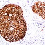
HER2 / ErbB2 / neu / CD340 Previously Observed Antibody Staining Patterns
Observed Subcellular, Organelle Specific Staining Data:
Anti-ERBB2 antibody staining is expected to be primarily localized to the cytosol and plasma membrane.
Observed Antibody Staining Data By Tissue Type:
Variations in HER2 / ErbB2 / neu / CD340 antibody staining intensity in immunohistochemistry on tissue sections are present across different anatomical locations. Low, but measureable presence of HER2 / ErbB2 / neu / CD340 could be seen inglandular cells in the fallopian tube, parathyroid gland, prostate, rectum, salivary gland, seminal vesicle, small intestine and thyroid gland, hematopoietic cells in the bone marrow, hepatocytes in liver, melanocytes in skin, pneumocytes in lung, squamous epithelial cells in the cervix, uterine, esophagus, tonsil and vagina and glandular cells in the fallopian tube, parathyroid gland, prostate, rectum, salivary gland, seminal vesicle and small intestine and thyroid gland. We were unable to detect HER2 / ErbB2 / neu / CD340 in other tissues. Disease states, inflammation, and other physiological changes can have a substantial impact on antibody staining patterns. These measurements were all taken in tissues deemed normal or from patients without known disease.
Observed Antibody Staining Data By Tissue Disease Status:
Tissues from cancer patients, for instance, have their own distinct pattern of HER2 / ErbB2 / neu / CD340 expression as measured by anti-HER2 / ErbB2 / neu / CD340 antibody immunohistochemical staining. The average level of expression by tumor is summarized in the table below. The variability row represents patient to patient variability in IHC staining.
| Sample Type | breast cancer | carcinoid | cervical cancer | colorectal cancer | endometrial cancer | glioma | head and neck cancer | liver cancer | lung cancer | lymphoma | melanoma | ovarian cancer | pancreatic cancer | prostate cancer | renal cancer | skin cancer | stomach cancer | testicular cancer | thyroid cancer | urothelial cancer |
|---|---|---|---|---|---|---|---|---|---|---|---|---|---|---|---|---|---|---|---|---|
| Signal Intensity | ++ | – | – | – | – | – | – | + | – | – | – | – | – | – | – | – | – | – | – | + |
| ERBB2 Variability | +++ | + | + | ++ | ++ | + | ++ | ++ | + | + | + | ++ | ++ | + | + | ++ | + | ++ | + | ++ |
| HER2 / ErbB2 / neu / CD340 General Information | |
|---|---|
| Alternate Names | |
| Receptor tyrosine-protein kinase erbB-2, CD340, cluster of differentiation 340, proto-oncogene Neu, Erbb2, ERBB2, Her2, HER2 | |
| Molecular Weight | |
| 185kDa | |
| Chromosomal Location | |
| 17q12 | |
| Curated Database and Bioinformatic Data | |
| Gene Symbol | ERBB2 |
| Entrez Gene ID | 2064 |
| Ensemble Gene ID | ENSG00000141736 |
| RefSeq Protein Accession(s) | NP_001005862, NP_001276866, NP_001276867, NP_004439, NP_001276865 |
| RefSeq mRNA Accession(s) | NR_110535, NM_001289937, NM_001289936, NM_001289938 NM_004448, NM_001005862 |
| RefSeq Genomic Accession(s) | NG_007503, NC_018928, NC_000017 |
| UniProt ID(s) | P04626, F5H1T4, X5DNK3, J3QLU9 |
| UniGene ID(s) | P04626, F5H1T4, X5DNK3, J3QLU9 |
| HGNC ID(s) | 3430 |
| Cosmic ID(s) | ERBB2 |
| KEGG Gene ID(s) | hsa:2064 |
| PharmGKB ID(s) | PA27844 |
| General Description of HER2 / ErbB2 / neu / CD340. | |
| This MAb is specific to ErbB2/HER2, shows minimal cross-reaction with other members of the family. ErbB2/HER2 is a member of the EGFR family. Receptors of this family are located on the plasma membrane, consist of an extracellular ligand-binding domain that is connected to a large intracellular domain by a single transmembrane sequence. ErbB2/HER2 protein is over-expressed in a variety of carcinomas especially those of breast, ovary. | |
-150x150.jpg)
There are no reviews yet.