
 PDF Datasheet
Human, Chimpanzee, Sheep, Rat, Mouse, and Chicken Anti-HSP27 Antibody Product Attributes
HSP27 Previously Observed Antibody Staining Patterns
Observed Subcellular, Organelle Specific Staining Data:
Anti-HSPB1 antibody staining is expected to be primarily localized to the cytosol and plasma membrane.
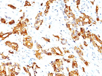
Observed Antibody Staining Data By Tissue Type:
Variations in HSP27 antibody staining intensity in immunohistochemistry on tissue sections are present across different anatomical locations. An intense signal was observed in decidual cells in the placenta, endothelial cells in the colon, epidermal cells in the skin, exocrine glandular cells in the pancreas, follicle cells in the ovary, glandular cells in the endometrium, epididymis, fallopian tube and gallbladder, keratinocytes in skin, Langerhans in skin, melanocytes in skin, myocytes in heart muscle, respiratory epithelial cells in the bronchus and nasopharynx, squamous epithelial cells in the esophagus, oral mucosa, tonsil and vagina, trophoblastic cells in the placenta and urothelial cells in the urinary bladder. More moderate antibody staining intensity was present in decidual cells in the placenta, endothelial cells in the colon, epidermal cells in the skin, exocrine glandular cells in the pancreas, follicle cells in the ovary, glandular cells in the endometrium, epididymis, fallopian tube and gallbladder, keratinocytes in skin, Langerhans in skin, melanocytes in skin, myocytes in heart muscle, respiratory epithelial cells in the bronchus and nasopharynx, squamous epithelial cells in the esophagus, oral mucosa, tonsil and vagina, trophoblastic cells in the placenta and urothelial cells in the urinary bladder. Low, but measureable presence of HSP27 could be seen inadipocytes in mesenchymal tissue, bile duct cells in the liver, fibroblasts in mesenchymal tissue, glandular cells in the breast and colon, hematopoietic cells in the bone marrow, hepatocytes in liver, islets of Langerhans in pancreas, Leydig cells in the testis and pneumocytes in lung. We were unable to detect HSP27 in other tissues. Disease states, inflammation, and other physiological changes can have a substantial impact on antibody staining patterns. These measurements were all taken in tissues deemed normal or from patients without known disease.
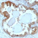
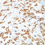
Observed Antibody Staining Data By Tissue Disease Status:
Tissues from cancer patients, for instance, have their own distinct pattern of HSP27 expression as measured by anti-HSP27 antibody immunohistochemical staining. The average level of expression by tumor is summarized in the table below. The variability row represents patient to patient variability in IHC staining.
| Sample Type | breast cancer | carcinoid | cervical cancer | colorectal cancer | endometrial cancer | glioma | head and neck cancer | liver cancer | lung cancer | lymphoma | melanoma | ovarian cancer | pancreatic cancer | prostate cancer | renal cancer | skin cancer | stomach cancer | testicular cancer | thyroid cancer | urothelial cancer |
|---|---|---|---|---|---|---|---|---|---|---|---|---|---|---|---|---|---|---|---|---|
| Signal Intensity | ++ | – | + | – | – | – | + | – | ++ | – | ++ | ++ | ++ | – | ++ | ++ | – | – | + | ++ |
| HSPB1 Variability | ++ | + | +++ | ++ | ++ | + | ++ | ++ | ++ | + | + | ++ | ++ | + | ++ | ++ | ++ | + | ++ | +++ |
| HSP27 General Information | |
|---|---|
| Alternate Names | |
| Heat shock protein 27, Hsp27, heat shock protein beta-1, HSPB1, HSPB1 , Hsp 27 | |
| Molecular Weight | |
| 27kDa | |
| Chromosomal Location | |
| 7q11.23 | |
| Curated Database and Bioinformatic Data | |
| Gene Symbol | HSPB1 |
| Entrez Gene ID | 3315 |
| Ensemble Gene ID | ENSG00000106211 |
| RefSeq Protein Accession(s) | NP_001531 |
| RefSeq mRNA Accession(s) | NM_001540, |
| RefSeq Genomic Accession(s) | NG_008995, NC_018918, NC_000007 |
| UniProt ID(s) | V9HW43, P04792 |
| UniGene ID(s) | V9HW43, P04792 |
| HGNC ID(s) | 5246 |
| Cosmic ID(s) | HSPB1 |
| KEGG Gene ID(s) | hsa:3315 |
| PharmGKB ID(s) | PA29511 |
| General Description of HSP27. | |
| It recognizes a 24-27kDa estrogen-regulated protein, identified as heat shock protein 27 (hsp27). Hsp27 was recently found to be identical to the estrogen-induced p29, 24K protein. About 50% of breast carcinomas are positive for hsp27 especially those that are also positive for estrogen,/or progesterone receptor. HSP27 has also been implicated in drug resistance in cancer cells. | |

There are no reviews yet.