
.jpg)
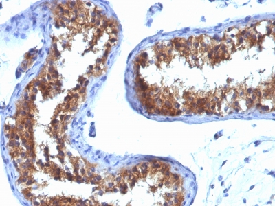
![Staining with Mouse monoclonal MVP [Clone 1032] antibody in formalin-fixed paraffin-embedded human testicular carcinoma.](https://enquirebio.com/wp-content/uploads/2019/01/MVP%20IHC%20human%20Testicular%20Carcinoma%20(1032).jpg)
 PDF Datasheet
Human Anti-Major Vault Protein Antibody Product Attributes
Major Vault Protein Previously Observed Antibody Staining Patterns
Observed Subcellular, Organelle Specific Staining Data:
Anti-MVP antibody staining is expected to be primarily localized to the cytosol.
Observed Antibody Staining Data By Tissue Type:
Variations in Major Vault Protein antibody staining intensity in immunohistochemistry on tissue sections are present across different anatomical locations. An intense signal was observed in glandular cells in the appendix, respiratory epithelial cells in the bronchus, glandular cells in the colon, peripheral nerve/ganglion in colon, glandular cells in the duodenum, macrophages in lung, respiratory epithelial cells in the nasopharynx, glandular cells in the rectum and small intestine, Leydig cells in the testis and urothelial cells in the urinary bladder. More moderate antibody staining intensity was present in glandular cells in the appendix, respiratory epithelial cells in the bronchus, glandular cells in the colon, peripheral nerve/ganglion in colon, glandular cells in the duodenum, macrophages in lung, respiratory epithelial cells in the nasopharynx, glandular cells in the rectum and small intestine, Leydig cells in the testis and urothelial cells in the urinary bladder. Low, but measureable presence of Major Vault Protein could be seen inhematopoietic cells in the bone marrow, Purkinje cells in the cerebellum, endothelial cells in the cerebral cortex, neuropil in cerebral cortex, glandular cells in the cervix, uterine, cells in the endometrial stroma in endometrium, glandular cells in the endometrium and epididymis, neuronal cells in the hippocampus, bile duct cells in the liver, germinal center cells in the lymph node, non-germinal center cells in the lymph node, exocrine glandular cells in the pancreas, glandular cells in the parathyroid gland, salivary gland and seminal vesicle, keratinocytes in skin, fibroblasts in mesenchymal tissue, peripheral nerve in mesenchymal tissue, cells in the white pulp in spleen and squamous epithelial cells in the tonsil and vagina. We were unable to detect Major Vault Protein in other tissues. Disease states, inflammation, and other physiological changes can have a substantial impact on antibody staining patterns. These measurements were all taken in tissues deemed normal or from patients without known disease.
Observed Antibody Staining Data By Tissue Disease Status:
Tissues from cancer patients, for instance, have their own distinct pattern of Major Vault Protein expression as measured by anti-Major Vault Protein antibody immunohistochemical staining. The average level of expression by tumor is summarized in the table below. The variability row represents patient to patient variability in IHC staining.
| Sample Type | breast cancer | carcinoid | cervical cancer | colorectal cancer | endometrial cancer | glioma | head and neck cancer | liver cancer | lung cancer | lymphoma | melanoma | ovarian cancer | pancreatic cancer | prostate cancer | renal cancer | skin cancer | stomach cancer | testicular cancer | thyroid cancer | urothelial cancer |
|---|---|---|---|---|---|---|---|---|---|---|---|---|---|---|---|---|---|---|---|---|
| Signal Intensity | ++ | – | + | ++ | ++ | – | ++ | ++ | – | + | + | + | ++ | – | ++ | – | ++ | – | ++ | + |
| MVP Variability | ++ | ++ | ++ | ++ | ++ | ++ | +++ | ++ | ++ | ++ | ++ | ++ | + | + | + | + | ++ | ++ | ++ | ++ |
| Major Vault Protein General Information | |
|---|---|
| Alternate Names | |
| Major vault protein, VAULT-1, VAULT1 | |
| Molecular Weight | |
| 104-110kDa | |
| Chromosomal Location | |
| 16p11.2 | |
| Curated Database and Bioinformatic Data | |
| Gene Symbol | MVP |
| Entrez Gene ID | 9961 |
| Ensemble Gene ID | ENSG00000013364 |
| RefSeq Protein Accession(s) | NP_001280134, NP_005106, NP_059447, NP_001280133 |
| RefSeq mRNA Accession(s) | NM_001293204, NM_001293205, NM_017458, NM_005115 |
| RefSeq Genomic Accession(s) | NC_000016, NC_018927 |
| UniProt ID(s) | X5D2M8, X5D7K9, Q14764, X5DNU0 |
| UniGene ID(s) | X5D2M8, X5D7K9, Q14764, X5DNU0 |
| HGNC ID(s) | 7531 |
| Cosmic ID(s) | MVP |
| KEGG Gene ID(s) | hsa:9961 |
| PharmGKB ID(s) | PA31332 |
| General Description of Major Vault Protein. | |
| Recognizes a protein of 104kDa-110kDa, characterized as major vault protein (MVP). Vaults are large ribonucleoprotein particles (RNPs) present in all eukaryotic cells. They have a complex morphology, including several small molecules of RNA, but a single protein species. The MVP accounts for >70% of their mass. Their shape is reminiscent of the nucleopore central plug. Treatment of cells with estradiol increases the amount of MVP in nuclear extract. The hormone-dependent interaction of vaults with ER is prevented in vitro by sodium molybdate. Antibodies to estrogen, progesterone, glucocorticoid receptors are able to co-immunoprecipitate the MVP. MVP is overexpressed in many neoplastic tissues, cell lines. Expression of MVP predicts a poor response to chemotherapy. | |
-150x150.jpg)
Reviews
There are no reviews yet.