
.jpg)
 PDF Datasheet
Human Anti-Napsin A Antibody Product Attributes
Napsin A Previously Observed Antibody Staining Patterns
Observed Antibody Staining Data By Tissue Type:
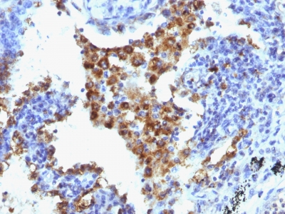
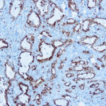
Variations in Napsin A antibody staining intensity in immunohistochemistry on tissue sections are present across different anatomical locations. An intense signal was observed in cells in the tubules in kidney and macrophages in lung. More moderate antibody staining intensity was present in cells in the tubules in kidney and macrophages in lung. Low, but measureable presence of Napsin A could be seen in. We were unable to detect Napsin A in other tissues. Disease states, inflammation, and other physiological changes can have a substantial impact on antibody staining patterns. These measurements were all taken in tissues deemed normal or from patients without known disease.
Observed Antibody Staining Data By Tissue Disease Status:
Tissues from cancer patients, for instance, have their own distinct pattern of Napsin A expression as measured by anti-Napsin A antibody immunohistochemical staining. The average level of expression by tumor is summarized in the table below. The variability row represents patient to patient variability in IHC staining.
| Sample Type | breast cancer | carcinoid | cervical cancer | colorectal cancer | endometrial cancer | glioma | head and neck cancer | liver cancer | lung cancer | lymphoma | melanoma | ovarian cancer | pancreatic cancer | prostate cancer | renal cancer | skin cancer | stomach cancer | testicular cancer | thyroid cancer | urothelial cancer |
|---|---|---|---|---|---|---|---|---|---|---|---|---|---|---|---|---|---|---|---|---|
| Signal Intensity | – | – | – | – | – | – | – | – | ++ | – | – | – | – | – | – | – | – | – | – | – |
| NAPSA Variability | + | + | + | + | + | + | ++ | + | ++ | + | + | + | + | + | ++ | + | + | + | + | + |
| Napsin A General Information | |
|---|---|
| Alternate Names | |
| Napsin-A, NAPSA | |
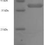
| Molecular Weight | |
| 37kDa | |
| Chromosomal Location | |
| 19q13.33 | |
| Curated Database and Bioinformatic Data | |
| Gene Symbol | NAPSA |
| Entrez Gene ID | 9476 |
| Ensemble Gene ID | ENSG00000131400 |
| RefSeq Protein Accession(s) | XP_011525842, NP_004842, XP_016883001 |
| RefSeq mRNA Accession(s) | NM_004851, XM_011527540, XM_017027512 |
| RefSeq Genomic Accession(s) | NC_018930, NC_000019 |
| UniProt ID(s) | O96009 |
| UniGene ID(s) | O96009 |
| HGNC ID(s) | 13395 |
| Cosmic ID(s) | NAPSA |
| KEGG Gene ID(s) | hsa:9476 |
| PharmGKB ID(s) | PA134891814 |
| General Description of Napsin A. | |
| Napsin is a pepsin-like aspartic proteinase connected with maturation of surfactant protein B.There are two closely related napsins, napsin A, napsin B. Napsin A is expressed as a single chain protein. Immunohistochemical studies revealed high expression levels of napsin A in human lung, kidney but low expression in spleen. Napsin A is expressed in type II pneumocytes, in adenocarcinomas of lung. The high specificity expression of napsin A in adenocarcinomas of lung is useful to distinguish primary lung adenocarcinomas from adenocarcinomas of other organs. | |
-150x150.jpg)
-150x150.jpg)
Reviews
There are no reviews yet.